SPLEEN
1. What are four of the functions of the spleen?
The spleen is the largest lymphoid organ of the body (Figure Q-20). It is unlike the lymph nodes in that the circulating fluid is blood instead of lymph.
It is the only organ specializing in filtering blood. A section cut from the spleen (Figure Q-21) shows that it is surrounded by a capsule that has connective tissue and smooth muscle cells. The amount of smooth muscle varies with species and is quite pronounced in carnivores. Trabeculae extend from the capsule that are composed of elastic fibers, collagen, and smooth muscle. Arteries, veins, lymph vessels, and nerves are contained within the trabeculae. The parenchyma (splenic pulp) of the spleen is composed of red and white pulp and is supported by the capsule, trabeculae, and reticular fibers. Most of the splenic pulp is red pulp and blood is held within the reticular fiber mesh, which represents the part of the spleen that acts as a filter; it has numerous fixed macrophages. The white pulp is lymphatic tissue distributed throughout the spleen as lymphatic nodules and sheaths of lymphatics around arteries and arterioles that produces lymphocytes. Blood enters the spleen from its hilus and from trabeculae and is distributed either to the lymph nodules via nodular capillaries or to the red pulp or venous sinuses via terminal capillaries. Blood entering the red pulp (reticular spaces) via the terminal capillaries is then able to enter the venous sinuses through slits in the venous sinus walls. Blood entering the reticular spaces provides for greater exposure to cells of the mononuclear phagocytic system (MPS). The venous sinuses collect filtered blood and drain into venules, and finally into trabecular veins.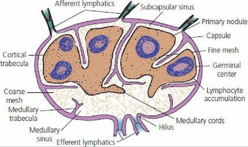
■ FIGURE 9-19 Internal structure of a lymph node. Lymph enters through afferent lymphatics and leaves through efferent lymphatics.
The lymph percolates through the coarse mesh, on which many fixed mononuclear phagocytic cells are located. Lymphocytes are produced in the primary nodules and accumulate throughout the fine mesh (dark tan). A fine mesh holds small lymphocytes better than a coarse mesh.

■ FIGURE 9-20 Projection of viscera on the left body wall of the female dog showing the location of the spleen relative to other body organs. The dog spleen is somewhat variable in position and its long axis can be almost longitudinal.
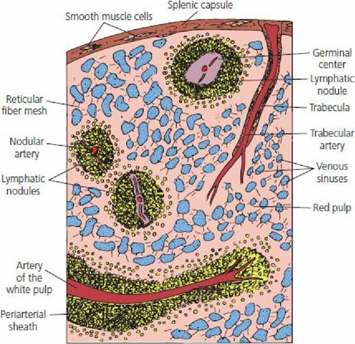
■ FIGURE 9-21 Schematic representation of the pig spleen. Multiple branches of the splenic artery enter the capsule and extend into the trabeculae. The lymphatic nodules and periarterial sheaths compose the white pulp that produces lymphocytes. The red pulp is the reticular fiber mesh that acts as a filter because of its fixed macrophages. Smooth muscle cells are present in the capsule and in the trabeculae. The venous sinuses collect filtered blood and drain into venules and finally trabecular veins (not shown).
Blood circulates through the spleen, and the spleen is active in the destruction of aged and abnormal erythrocytes by the numerous MPS cells. Also, the spleen is a storage depot of iron obtained from the destruction of erythrocytes. The spleen is an important reservoir of blood, especially of red blood cells, which accumulate within the venous sinusoids. Contraction of the spleen is possible because of the smooth muscle and occurs when more red blood cells are needed. Splenic contraction that accompanies excitement in the dog can increase the packed cell volume from 40% to more than 50%.
■