Cardiac Contractions Are Initiated by Action Potentials That Arise Spontaneously in Specialized Pacemaker Ceils
Because cardiac muscle tissue forms a functional syncytium, and because a cardiac action potential leads to contraction, any one cardiac muscle cell can initiate a heartbeat. In other words, if a single cardiac muscle cell depolarizes to threshold and forms an action potential, that action potential will propagate from cell to cell, throughout the heart, and cause the whole heart to contract.
Most cardiac muscle cells have the property of remaining stable at a resting membrane potential; they never form action potentials by themselves. However, a few specialized cardiac muscle cells have the property of depolarizing spontaneously toward the threshold for the formation of action potentials. When any one of these specialized cells reaches threshold and forms an action potential, a heartbeat results. Cardiac cells that depolarize spontaneously toward threshold are called pacemaker cells because they initiate heartbeats and therefore determine the rate, or pace, of the heart.Although all spontaneously depolarizing cells in the heart are called pacemaker cells, only one pacemaker cell, the one that reaches threshold first, actually triggers a particular heartbeat. In the normal heart, the pacemaker cells that depolarize most quickly to threshold are located in the sinoatrial (SA) node. The SA node is in the right atrial wall, at the point where the venae cavae enter the right atrium.
Because it has spontaneously depolarizing pacemaker cells, the heart initiates its own muscle action potentials and contractions. Motor neurons are not necessary for initiating cardiac contractions, whereas they are needed for initiating skeletal muscle contractions. Motor neurons (sympathetic and parasympathetic) do affect the heart rate, by influencing the rapidity of the pacemaker cells' depolarization to threshold level, but the heart continues to beat even without any sympathetic or parasympathetic influences. Thus a denervated heart still beats, whereas a denervated skeletal muscle remains relaxed (in fact, paralyzed). The ability of the heart to beat without neural input enables surgically transplanted hearts to function. When a donor’s heart is connected to a recipient's circulation during cardiac transplantation, no nerves are attached to the heart. The pacemaker cells in the transplanted
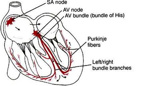
FIGURE 19-2 Specialized conduction system of the heart is responsible for the initiation and organization of cardiac contractions.The system is composed of specialized cardiac muscle fibers, not nerves. AVf Atrioventricular; SAf sinoatrial.
heart initiate its action potentials and contractions, just as they do in a normal heart. The only factor missing is control of the heart rate through cardiac sympathetic and parasympathetic nerves.