A System of Specialized Cardiac Muscle Cells Initiates and Organizes Each Heartbeat
Each normal heartbeat is initiated by an action potential that arises spontaneously in one of the pacemaker cells in the SA node (Figure 19-2). Once formed, the action potential propagates rapidly, from cell to cell, across the right and left atria, causing both atria to contract.
Next, the action potential works its way, from cell to cell, through a special pathway of cardiac muscle cells that lies between the atria and the ventricles. This pathway consists of the atrioventricular (AV) node and the first part of the AVz bundle, also called the bundle of His. The AV node and AV bundle provide the only route for the conduction of action potentials from the atria to the ventricles. Elsewhere, the atria and ventricles are separated by a layer of connective tissue, which can neither form nor propagate action potentials. In addition to providing the only conductive pathway between the atria and the ventricles, the AV node and the first part of the AV bundle have the special property of very slow conduction of action potentials. It takes 50 to 150 msec for an atrial action potential to travel through the AV node and the first part of the AV bundle; that is, it takes 50 to 150 msec for an atrial action potential to propagate into the ventricles. Slow conduction through the AV junction creates the delay between atrial and ventricular contractions.Once past the slowly conducting ceils of the AV junction, the cardiac action potential enters a branching network of specialized cardiac cells that have the property of extremely rapid conduction of action potentials from cell to cell. This network begins with the AV bundle, which has slowly conducting cells in its first portion (connected to the AV node) and rapidly conducting cells beyond that. The AV bundle divides to form the rapidly conducting left and right bundle branches. At the ventricular apex, the bundle branches break up into a dispersed network of Purkinje fibers, which carry the action potential rapidly along the inner walls of both ventricles.
The Purkinje fibers propagate action potentials into the normal ventricular muscle fibers within the inner walls (subendocardial layers) of both ventricles. From there, the action potentials propagate quite rapidly outward, from cell to cell, through the ventricular walls. As the action potential reaches each ventricular muscle fiber, that fiber contracts. The extremely rapid conduction of the cardiac action potential, from cell to cell, through the latter portion of the AV bundle, the bundle branches, and the Purkinje system results in a nearly synchronous contraction of all the fibers in both ventricles.The SA and AV nodes, AV bundle, bundle branches, and Purkinje fibers are collectively called the specialized conduction system of the heart. This system is composed of specialized cardiac muscle cells, not nerves. The particular characteristics of the components in the specialized conduction system cause each heartbeat to follow a specific, patterned sequence. In a normal beat, both atria contract, almost simultaneously. Next, there is a brief pause (caused by slow conduction of the action potential through the AV node). The two ventricles then contract, almost simultaneously. Finally, the entire heart relaxes and refills.
Figure 19-3 reemphasizes the role of the specialized conduction system in initialing and organizing a normal cardiac contraction. In this "time lapse" illustration, atrial excitation begins at time t = O, when one SA node cell has reached threshold and an action potential is just beginning to propagate out of the SA node and into regular atrial tissue. Within O. I second, the action potential has propagated completely across the right and left atria, and a coordinated contraction of both atria is just beginning. As Iheaction potential propagates across the atria, it also depolarizes the first cells in the AV node, beginning at time t = 0.04 second. While the atria are in a depolarized (excited) state, the action potential is propagating slowly from cell to cell through the AV node and first part of the AV bundle.
After traversing this slowly conducting region, the action potential is propagated rapidly through the remainder of the bundle of His and its branches. The action potential arrives at the ventricular apex at time t = 0.17 second. Note that it takes about 0.13 second (0.17 - 0.04 second) for the action potential to travel through the AV node and bundles; that is, 0.13 second represents a typical delay between atrial depolarization and ventricular depolarization. From the ventricular apex, the action potential is propagated rapidly throughout both ventricles by Purkinje fibers. Ventricular excitation (depolarization) is complete by time t = 0.22 second, and both ventricles contract. By this time the atria have repolarized to a resting state and are relaxing. After ventricular excitation and contraction, the ventricles relax, and the whole heart remains in a resting state until the next beat is originated by an SA node pacemaker cell.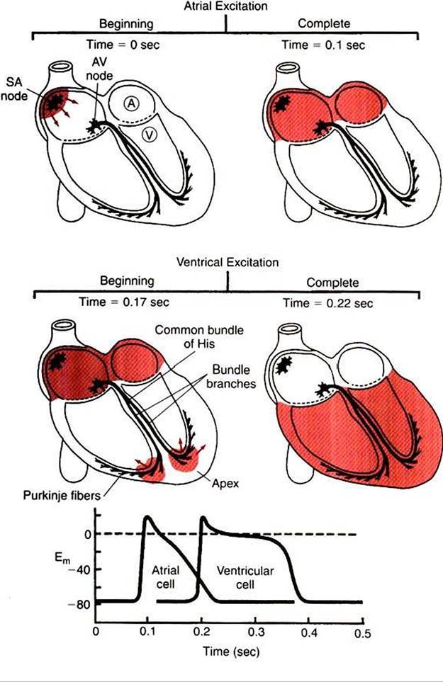
FIGURE 19-3 Heart is pictured at four instants during initiation of a normal contraction. Top left (time t= 0 sec), Pacemaker cell in the sinoatrial (SA) node has just reached threshold, and an action potential has begun to propagate outward across the atria. Top right (t= 0.1 sec), Action potential has reached all parts of both atria (all atrial cells are at the plateau of their action potentials). Middle left (t = 0.17 sec), Action potential has passed through the atrioventricular (AV) node and down the bundle branches and has just reached the ventricular apex. Middle right (t = 0.22 sec). Action potential has just finished propagating outward through the walls of both ventricles (all ventricular cells are at the plateau of their action potentials). Bottom, Graph shows the timing of action potentials in a left atrial cell (labeled A, top left) and a left ventricular cell (V, top left). Their locations make these among the last atrial and ventricular cells to be depolarized as action potentials propagate across the atria and ventricles, respectively. Em, Membrane potential in millivolts.