Cardiac Cycle
The cardiac cycle is one complete cycle of cardiac contraction and relaxation (heartbeat). The events of the cardiac cycle occur in a specific sequence, and for descriptive purposes, the continuous cycle is divided into phases or periods marked by different events.
Figure 182 illustrates the changes in blood pressures and volumes in the left atrium and left ventricle and pressure changes in the aorta during the cardiac cycle, and it identifies some phases of the cycle. The sequence of changes in blood pressures and volumes in the right atrium, right ventricle, and pulmonary trunk are similar and occur in the same sequence, but the magnitude of pressure changes in the right ventricle and pulmonary trunk are much lower. The unique characteristics of the pulmonary circulation are discussed in Chapter 19.Diastole (dilation, from the Greek dia, apart; stello, place or put) refers to the relaxation of a chamber of the heart just prior to and during the filling of that chamber. Systole (contraction, from Gr. syn, together; stello, place) refers to the contraction of a chamber of the heart that drives blood out of the chamber. The adjectives atrial and ventricular can be used with diastole or systole to describe the activity of specific cardiac chambers (e.g., atrial systole refers to atrial contraction in Figure 18-2). However, when the cardiac cycle is divided into only two general phases (diastole and systole) without specifying a chamber, it is generally assumed that division is based on ventricular activity. Thus, when the heart is said to be in systole or the systolic phase of the cardiac cycle, it is usually understood that this refers to ventricular systole. (Note in Figure 18-2 the atrial contracts while the ventricle remains relaxed, i.e., ventricular diastole.)
Two distinct heart sounds can be heard during each cardiac cycle in all domestic species, and these are typically described as lub (first sound, or S1) and dub (second sound, or S2).
These sounds are separated by a short interval and followed by a longer pause (Figure 18-2). The pause increases with slower heart rates. These sounds are used clinically to divide the cardiac cycle into two phases, systole (ventricular) and diastole (ventricular). The first sound marks the beginning of systole, and the second sound marks the beginning of diastole (Fig. 18-2).Systole
As the ventricles begin their contraction, blood pressure increases in them. Almost immediately, the pressure within each ventricle exceeds the pressure within their respective atria, and the pressure differences force the A-V valves closed. The first heart sound is associated with closure of the right and left A-V valves (Fig. 18-2).
The ventricles continue to contract and pressure continues to increase during the early part of systole. At this point in the cycle, all four heart valves are closed, and all remain closed until pressure in the ventricles exceeds that in the arterial vessels that they supply (aorta for left ventricle and pulmonary trunk for right ventricle). The period of systole during which all valves are closed is the isovolumetric
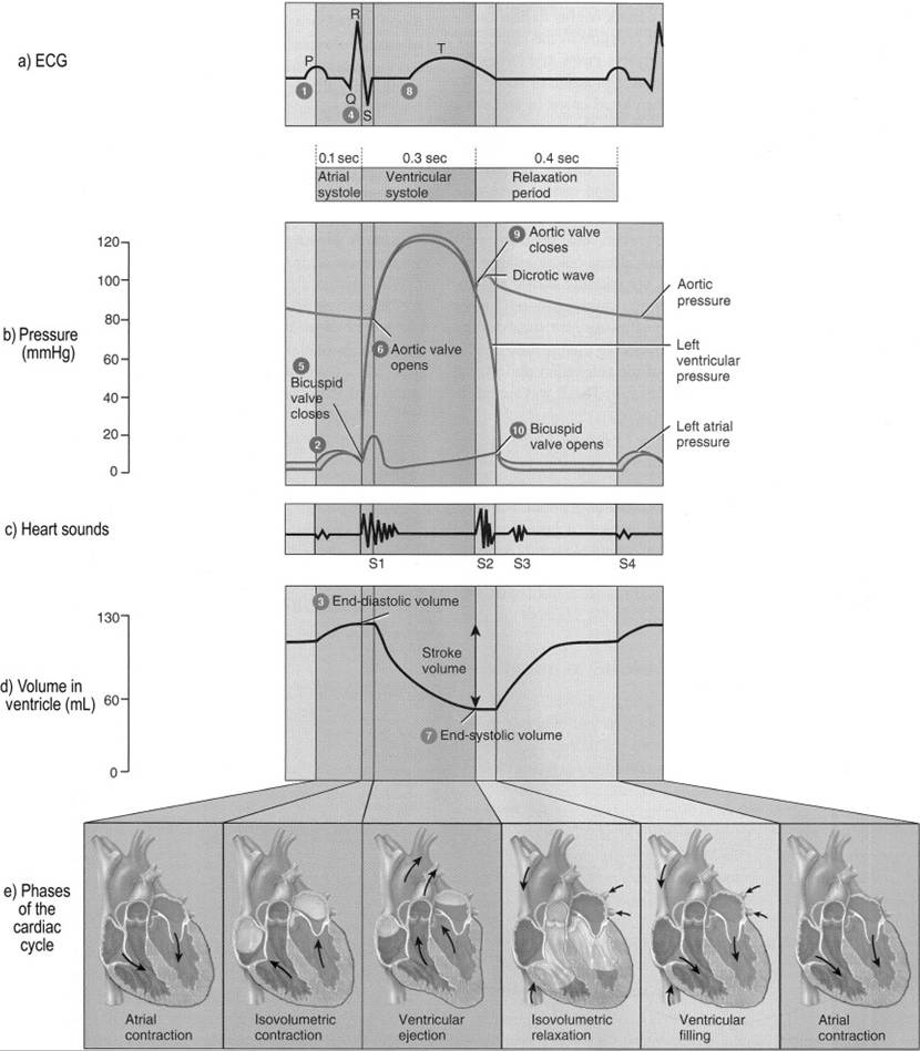
Figure 18-2. Timing of various events of the cardiac cycle. Pressures, volumes, and times represent typical values for an adult human. These characteristics would be different among domestic animals of variable sizes and with different heart rates. However, the sequence of the events and the relative differences in pressures between the atrium, ventricle, and aorta would be the same. Note that the complete period for ventricular diastole would include the segment in this diagram labeled as atrial contraction. (Reprinted with permission from Tortora, G.J. and Derrickson, B. Principles of Anatomy and Physiology, 11th ed. Hoboken, NJ: John Wiley & Sons, 2006.)
contraction period, because during it the volume of each ventricle remains constant (Fig.
18-2).When ventricular pressures exceed those in their respective arterial vessels, the semilunar valves open to permit ejection of blood. There is no sound associated with the opening of the semilunar valves. An initial rapid ejection phase of systole is followed by a reduced ejection phase, during which ventricular and arterial pressures fall (Fig. 18-2).
The elasticity of the aorta and pulmonary trunk maintains blood pressure in these vessels even though the ventricles begin to relax. When the blood pressures in these vessels are greater than the pressures in their associated ventricles, the pressure differences close the semilunar valves. The second heart sound is associated with closure of the aortic and pulmonary valves, and this sound is used to mark the end of systole (ventricular) and the beginning of diastole (ventricular) (Fig. 18-2).
Stroke volume is the volume of blood ejected from each ventricle during a single cardiac cycle. Normally, the stroke volumes for the right and left ventricles are the same. if these are not equal, blood tends to accumulate in either the systemic or pulmonary circulation.
Diastole
At the beginning of diastole, both the semilunar and A-V valves are closed, so the initial phase is termed isovolumetric relaxation. When ventricular relaxation reaches the point that atrial blood pressures exceed ventricular blood pressures, the pressure differences open the A-V valves. While the A-V valves are closed during systole and early diastole, blood continues to flow into the right and left atria from the systemic and pulmonary circulations, respectively. The accumulation of blood within the atria increases atrial blood pressure. When A-V valves open, much of the accumulated blood flows rapidly into the ventricles. Most ventricular filling occurs during this period prior to any atrial contraction (Fig. 18-2).
Blood continues to flow into the atria throughout diastole, and because the A-V valves are open, blood flows directly through the atria into the ventricles.
As mentioned earlier, diastole is the phase of the cardiac cycle that lengthens most with slow heart rates, so slow heart rates provide a long period for ventricular filling.Atrial contraction (atrial systole) occurs during ventricular diastole, forcing an additional volume of blood into the ventricles, but this amount is relatively small (perhaps 15%) compared to the volume already in the ventricles.
The volume of blood in each ventricle at the end of diastole is the end-diastolic volume (EDV). During systole, each ventricle ejects only a percentage of its EDV (Fig. 18-2), typically 40-60%. The percentage of the EDV that is ejected is the ejection fraction.
Heart Sounds and Murmurs
The first and second heart sounds are associated with valve closures, but turbulent blood flow and vibrations of large vessels induced by the closures are believed to be the actual causes of the sounds. Third and fourth heart sounds may be heard in some normal horses and cattle with relatively slow heart rates. The third sound is associated with the rapid ventricular filling phase after the initial opening of the A-V valves, and the fourth sound is associated with atrial contractions (Fig. 18-2).
Heart murmur is a general term for any abnormal heart sound. Abnormalities in heart valves are a common cause of murmurs. Murmurs may occur when a valve fails to close completely (valvular insufficiency) and blood flow goes in the wrong direction at the wrong time. Murmurs may also occur when a valve fails to open completely (valvular stenosis) and blood is forced through a smaller than normal opening.
Echocardiography
Echocardiography is the use of ultrasound to image the heart and associated structures. it is a noninvasive procedure that provides dynamic images permitting visualization of structures in the heart as it contracts and relaxes. This provides much more information on cardiac function than can be obtained with a single static image, such as a radiograph.