Electrical Activity of the Heart
Like skeletal muscle, contraction of each cardiac muscle cell requires an action potential on the cell membrane (Fig. 18-3A), and the action potential brings about the release of calcium from intracellular stores.
The calcium binds to regulatory proteins on the thin filaments, and movement of the regulatory proteins permits an interaction between actin and myosin to bring about contraction (see Chapter 8). However, unlike skeletal muscle, each cardiac muscle cell is not innervated by a motor neuron that is responsible for eliciting an initial action potential. in the heart, the initial action potential occurs spontaneously in a specialized group of myocardial cells found in the sinoatrial (SA) node of the heart (Fig. 18-4), from which the action potential is propagated around the heart to bring about contraction of all cardiac muscle cells. The cell-to-cell propagation in the heart is possible because the intercalated disks provide an electrical connection between myocardial cells (Fig. 1-6C).Sinoatrial Node and Heart Rate
Figure 18-3B shows a series of action potentials from SA node cells. A unique feature of the electrical activity of these cells is that the resting membrane potential is unstable. This instability permits SA node cells to depolarize spontaneously to threshold, where an action potential is generated. The SA node (Fig. 18-4) is termed the pacemaker of the heart because each action potential that spontaneously develops in the SA node is propagated around the heart to stimulate action potentials in all myocardial cells and produce a contraction.
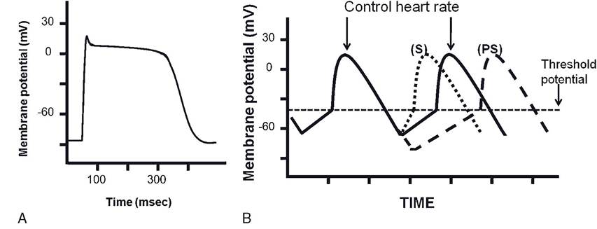
Figure 18-3A. Action potential of cardiac muscle contractile cell. Electrically gated calcium channels in cell membrane are open during the prolonged plateau phase of the action potential to permit calcium ions to enter from the extracellular fluid.
The entering calcium stimulates the release of more calcium from the sarcoplasmic reticulum. The initial depolarization phase involves electrically gated sodium channels, and potassium channels are involved with membrane repolarization.Figure 18-3B Four sinoatrial cell membrane potentials and action potentials to illustrate effects of sympathetic (S) and parasympathetic (PS) stimulation on a control heart rate. Sympathetic stimulation reduces the time period before the next action potential to increase heart rate. Parasympathetic stimulation prolongs the time period before the next action potential to reduce heart rate.
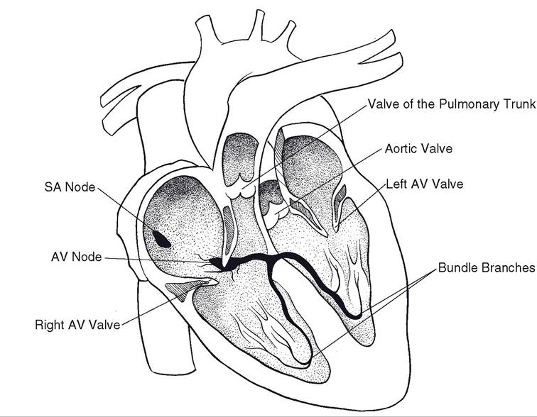
Figure 18-4. Impulse generation and conduction system of the mammalian heart.
Both sympathetic and parasympathetic nerves innervate the SA node. By their actions on cells in the sA node, sympathetic nerves increase the rate of spontaneous action potentials and parasympathetic nerves reduce the rate (Fig. 18-3B). This is the means by which sympathetic stimulation increases heart rate and parasympathetic stimulation reduces heart rate. Parasympathetic nerves continuously inhibit the SA node in the heart of a resting animal, and this constant inhibition is responsible for the resting heart rate. During light exercise or with excitement, the parasympathetic inhibition is first reduced to permit an increase in heart rate. With greater excitement or more intense exercise, sympathetic stimulation increases, further increasing heart rate. Highly trained athletic animals, such as racing Thoroughbreds, have relatively high levels of parasympathetic stimulation to their hearts at rest.
Atrioventricular Node and Other Specialized Conductive Cells in the Heart
The atrioventricular node (A-V node) and the common bundle, or bundle of His, are also myocardial cells specialized for conducting action potentials. The A-V node is in the intra- atrial septum, and the common bundle extends from the A-V node into the ventricle (Fig.
18-4) through the fibrous connective tissue of the cardiac skeleton. The cardiac skeleton separates the cardiac muscle of the atria and ventricles, so the only direct electrical connection is through the A-V node and common bundle. The common bundle divides into several branches that rapidly propagate action potentials throughout the ventricle (Fig. 18-4). The individual cells that make up these branches are the Purkinje fi bers.Cells of the A-V node are specialized to conduct action potentials more slowly than other myocardial cells. This characteristic allows enough time for the atria to depolarize completely and contract before action potentials spread into the ventricles to stimulate their contraction. The atrial contraction completes the filling of the ventricles so that ventricular contraction can eject a larger volume. The slow conduction through the A-V node is A-V node delay. sympathetic and parasympathetic nerves that increase and reduce conduction velocity, respectively, also innervate the A-V node.
Electrocardiography and Arrhythmias
Electrocardiography is the recording of electrical activity on the surface of the body that reflects the electrical activity in the heart. Recording electrodes are placed on the surface of the body at specific sites, and the recorded electrical activity reflects the summated electrical activity of the heart. Because of the specificity of the sites for the placement of electrodes, the patterns of electrical activity associated with a cardiac cycle are predictable, and comparisons can be made between animals. The lead is a specific combination of sites where the recording electrodes are placed on the body. An electrocardiogram (ECG) is the actual recording.
Figure 18-5 shows a typical lead II (or limb lead ii) electrocardiogram recorded from a dog. Major waves are P, Q, R, S, and T. The P wave is associated with atrial depolarization. The QRs complex is associated with ventricular depolarization, and the T wave is associated with ventricular repolarization.
The period between the P and Q waves is associated with A-V node delay. On a typical recording from a normal dog, the waves range from 1 to 2 mV and 0.2 to 0.3 seconds. Differences in wave shape and size among species are due to normal differences in the pattern of conduction of action potentials around the heart.Arrhythmia is a general term for any abnormality in cardiac electrical activity, including rate, rhythm, and the propagation
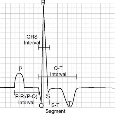
Figure 18-5. Typical canine lead II electrocardiogram.
of action potentials around the heart. Some apparently normal, healthy animals have a high incidence of cardiac arrhythmias in certain conditions. For example, it is fairly common for racing Thoroughbreds to have an apparent abnormality in A-V node conduction at rest. This is characterized by a reduction in or inhibition of the conduction of action potentials through the A-V node. These disappear with exercise. A likely cause is a relatively high parasympathetic neural input to the heart at rest that damps A-V node conduction. The relatively high parasympathetic input is normally reduced with exercise.