CARDIAC VESSELS AND NERVES
The heart is lavishly supplied with blood, receiving about 15% of the output of the left ventricle. The supply
is led through the coronary arteries that spring from two of the three sinuses above the semilunar cusps at the beginning of the aorta (Figure 7-17).
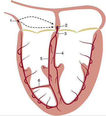
The left coronary artery is usually the larger. It arises above the caudosinistral cusp and reaches the coronary groove by passing between the left auricle and the pulmonary trunk; it divides almost at once. The left (para- conal) interventricular branch follows the like-named groove toward the apex of the heart (Figure 7-18Z2'). The trunk continues as a circumflex branch (Figure 7-18Z2") that follows the coronary groove toward the caudal aspect of the heart, where it may terminate close to the origin of the right (subsinuosal) interventricular groove (horse and pig) or continue into this (carnivores and ruminants) (Figure 7-19, A-B, and Figure 7-20).
The right coronary artery arises above the cranial cusp (Figure 7-17Z6) and reaches the coronary groove after passage between the right auricle and pulmonary trunk. It pursues a circumflex course that either fades toward the origin of the subsinuosal groove or turns into it in those species in which the left artery has the restricted distribution. Both coronary arteries send other branches, of varying size and constancy of position, to neighboring parts of the atrial and ventricular
Figure 7-14 Schematic drawing of the conducting system of the heart. The broken lines suggest the passage of the excitation wave through the atrial wall. 1, Sinuatrial node; 2, atrioventricular node; 3, atrioventricular bundle; 4, left limb; 5, right limb; 6, branch of right limb traversing the septomarginal band.
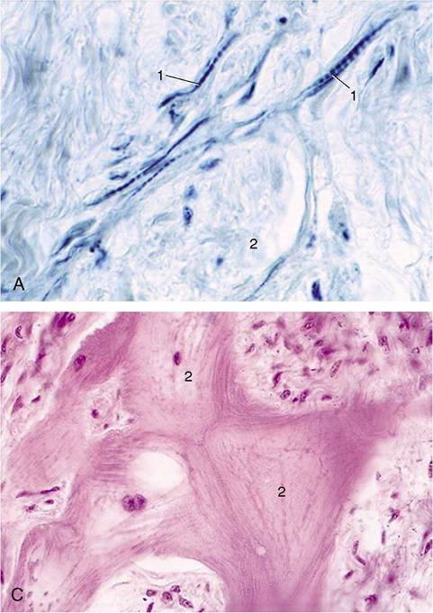
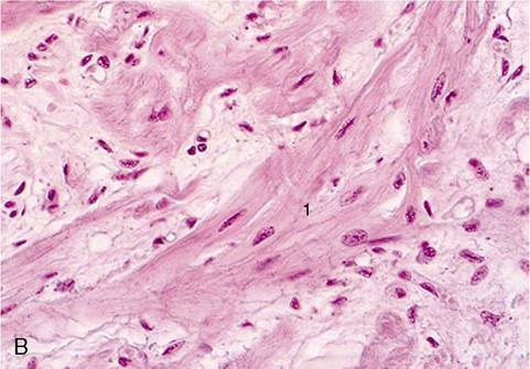
Figure 7-15 A, Sinuatrial node of the equine heart.
1, Nodal myofibers; 2, bundle of nerve fibers (I-HE) (279?). B and C, Atrioventricular node of equine heart (HE) (279?). 1, Nodal myofibers; 2, Purkinje cells with abundant glycogen.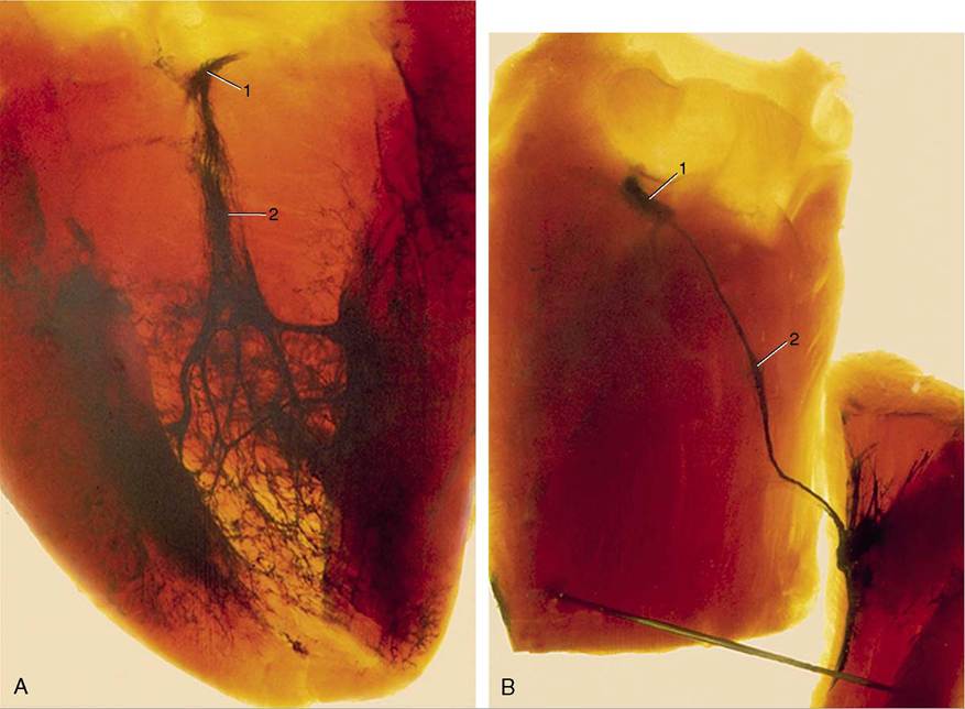
Figure 7-16 A, Cleared specimen of left ventricle. 1, Atrioventricular node; 2, left crus of atrioventricular trunk (injected blue). B, Cleared specimen of right ventricle. 1, Atrioventricular node; 2, right crus of the atrioventricular trunk, continuing into the moderator band.
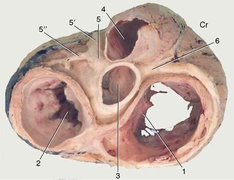
Figure 7-17 Dorsal view of the base of the heart after removal of the atria. The coronary arteries are exposed. 1, Right atrioventricular valve; 2, left atrioventricular valve; 3, aortic valve; 4, pulmonary valve; 5, left coronary artery; 5', paraconal interventricular branch; 5", circumflex branch; 6, right coronary artery. Cr, cranial.
walls. Very small twigs extend some distance into the cores of the valve cusps (Figure 7-21).
Anastomoses are not formed between the main branches of the coronary arteries but are numerous between the lesser branches. Even so, sudden closure of one of these small vessels cannot usually be compensated; it leads to local infarction of the cardiac muscle.
Blood is principally returned to the heart through the great cardiac vein that opens separately into the right atrium via the coronary sinus (Figure 7-19/3,4). Rather surprisingly, many very small (thebesian) veins open directly into all four heart chambers.
The innervation of the heart is complicated topographically, but happily the details mainly concern physiologists. A sympathetic contribution is routed through the caudal cervical and first few thoracic ganglia of the sympathetic trunk. The postganglionic fibers form cardiac plexuses within the cranial mediastinum before extending to the heart wall (Figure 7-22). Parasympathetic fibers branch from the vagus nerves, either directly or after short passage within the recurrent laryngeal nerves. They end on nerve cells in the
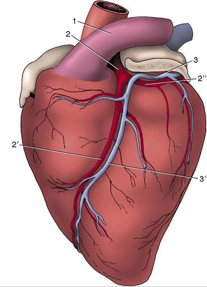
Figure 7-18 Branching of the left coronary artery of the heart, viewed from the left. The left auricle has been shortened. 1, Pulmonary trunk; 2, left coronary artery; 2', paraconal interventricular branch; 2", circumflex branch; 3, great cardiac vein (continued by the coronary sinus on the right side of the heart); 3', paraconal interventricular tributary of 3.
heart wall, especially within and about the sinuatrial and atrioventricular nodes. Many of the postganglionic fibers pass to the nodes, but others reach the periphery of the heart by following the atrioventricular bundle and its branches.