CARDIOVASCULAR SYSTEM
The heart has three chambers: two atria and one ventricle, and lies cranially at the level of the pectoral girdle in most lizards (Fig. 4.15). In the more advanced lizards like monitors (Varanus spp.) the heart has descended caudally to lie in the middle of the thoracoabdominal cavity.
Paired right and left aorta fuse caudal to it to form the dorsal aorta. A large ventral abdominal vein lies along the inner surface of the midline so this must be avoided when making a celiotomy incision (Barten 1996; Bennett & Mader 1996). A renal portal system is present, as in all reptiles.Larger lizards (and crocodiles) have a vasovagal reflex whereby pressure on the eyeballs decreases the heart rate and blood pressure. This can be used by clinicians to perform non-painful procedures like radiography (Bennett, RA 1996).
CLINICAL NOTE
Incise paramedian or alternatively incise the linea alba with caution as the ventral abdominal vein is suspended in the broad ligament and is only a few millimeters away. Keep it protected with saline gauze during the procedure.
Venepuncture sites
(See Murray 2000; Redrobe & MacDonald 1999.)
• Ventral coccygeal vein - The vein must be accessed caudal to the cloaca to avoid damaging the hemipenes in males. Access is usually about one third of the way from the vent and the needle is inserted ventrally in the midline. In some species a lateral approach may be used by inserting the needle perpendicular to the tail and ventral to the lateral vertebral processes (Murray 2000).
• Cardiac - as the heart cannot be held this is not as safe in lizards as in snakes.
• Axillary venous plexus - this is located near the shoulder joint at the caudal aspect of the humerus. Lymph dilution can occur.
• Ventral abdominal vein - this vein is very fragile so anesthesia is advisable to avoid lacerating it. The vein is approached two thirds of the distance caudally on the midline where it lies superficially just under the skin and muscle and is useful for lizards, like geckos,
with short tails (Murray 2000).
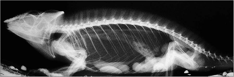
Figure 4.15 • Lateral (horizontal beam) radiograph of Panther chameleon (Furcifer pardalis). The heart lies at the pectoral inlet in most lizards. More advanced lizards like the monitors (Varanus spp.) have the heart lying more caudally in mid celom.