SKELETAL SYSTEM
Skull
In most species (except burrowing lizards and skinks) the head is narrower than the body. Like snakes, lizards have a kinetic skull which is thought to enable the upper and lower jaw to be closed simultaneously over prey (Figs.
4.6 and 4.7). The lower jaw is further increased in gape by a condition known as streptostyly. This is when the quadrate bone has no firm connection (owing to the absence of the temporal arch) and can move backward and forward. The main advantage of this is that it gives the adductor muscles that close the jaw a better mechanical advantage when biting (Bellairs 1969a; King 1996b).Although lizards have a large gape a united mandibular symphysis means they cannot open their mouths as wide as snakes. They compensate by having stronger jaws to help them immobilize, crush or tear at prey.
The adductor muscles extend from the temporal region to the lower jaw (Fig. 4.8). The main adductor muscle is the pterygoideus, which arises from the pterygoid bones on the palate and inserts on the caudal aspect of the lower jaw where it forms a large belly of muscle. It is this muscle that
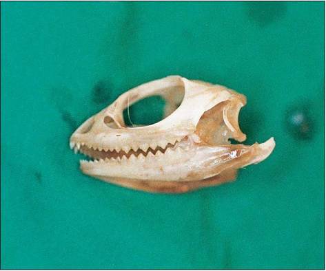
Figure 4.6 • Skull of juvenile bearded dragon (Pogona vitticeps).
can give the heavy jowled appearance to male lizards. The depressor mandible, which opens the jaw, arises from the back of the skull and inserts on the retroarticular process of the mandible. It is much weaker than the muscles that close the jaw (Bellairs 1969b; King 1996a).
CLINICAL NOTE
Always use a mouth gag when examining the mouths of large healthy lizards as the jaw can close like a trapdoor causing considerable damage and pain to unwary fingers.
Lizards are very mobile, having a flexible backbone, well- developed legs, and a long tail for counterbalance.
All the vertebrae except the cervical ones bear ribs, leaving little flank area. Ventrally the ribs either join the sternum, the opposite member, or end free in the body wall. The number of tail vertebrae is usually higher than the number of presacral vertebrae.More primitive lizards and tortoises have short limbs directed sideways, giving them their characteristic swinging gait. Advanced lizards have rotated these limbs towards the body so that the elbow faces caudally and the knee cranially. This form of limb orientation creates limbs that act as better shock absorbers.
Pectoral girdle and forelimbs
The pectoral girdle is composed of the scapula, coracoid bone and clavicle, and often an interclavicle. The forelimb has a short humerus and radius/ulna, with two rows of carpal bones. Both fore and hind feet are pentadactyl and the number of phalanges follows the formula 2,3,4,5,3 (from thumb to fifth digit), which gives rise to an asymmetrical foot (Fig. 4.9).
Pelvic girdle and hindlimbs
The pelvic girdle consists of a caudodorsally directed ilium, ischium and pubis and is firmly braced against the sacrum. The hindlimb is longer than the forelimb, owing to the elongated femur and phalanges. The tarsal bones have fused to form two bones called the astragalus-calcaneum, which articulates with the tibia and fibula. Flexion occurs between this joint and the rest of the foot. The hind foot has the first four metatarsal bones lying together while the fifth metatarsal lies separated with a backward-pointing hook. This allows the fifth digit to be opposed to the first, giving a better grip (Bellairs 1969a; King & Custance 1982; Pough 1998d) (Fig. 4.10).
Burrowing lizards have lost their limbs but, unlike snakes, they still retain their pectoral and pelvic girdle. Some lizards
Reptiles
61
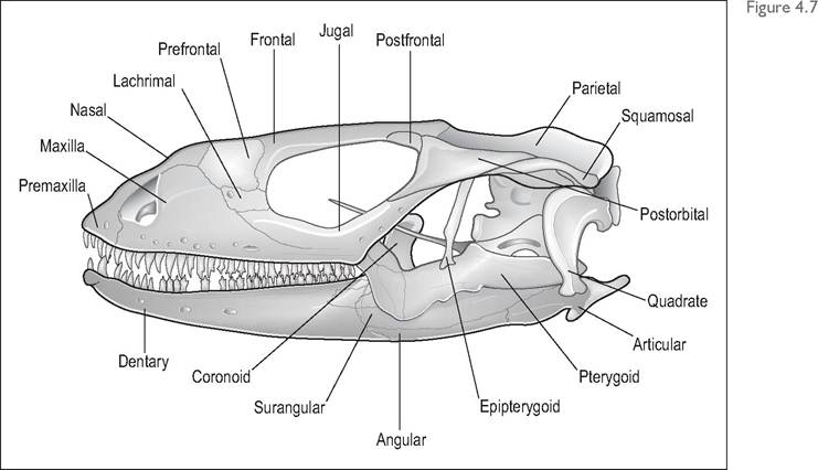
Skull of lizard.
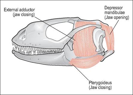
Figure 4.8 • Diagram of lizard skull showing main adductor (jaw closing) muscles.
As the adductor muscles are extremely strong a mouth gag should be used when handling large lizards.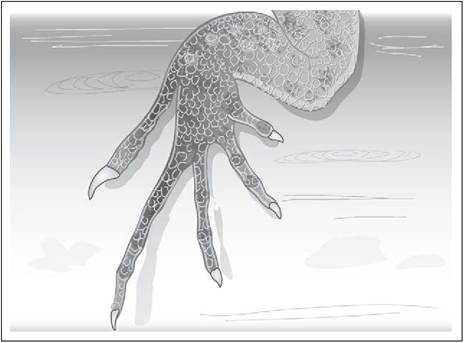
Figure 4.10 • Right hind foot of lizard showing first four metatarsals and separately attached fifth digit.
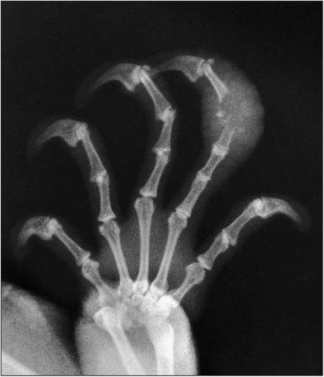
Figure 4.9 • Radiograph of right forefoot of Green iguana (iguana iguana) showing osteomyelitis at phalanges 3—4 of fourth digit. Both fore- and hind feet are pentadactyl in lizards with the number of phalanges following the formula 2,3,4,5,3 from thumb to fifth digit.
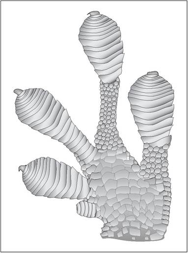
Figure 4.1 1 • Ventral view of gecko foot showing subdigital adhesive lamellae. These enable geckos to walk upside down on smooth surfaces.
GENERAL INTEREST
can hold their forelegs off the ground and run along on their hindlimbs in bipedal motion. Such species generally have a long tail as a counterbalance and lightweight thigh muscles.
Feet often have specialized adaptations. Many geckos have adhesive lamellae on their digits that allow them to walk on smooth vertical surfaces (Fig. 4.11). Chameleons have pincer-like zygodactyl feet with the first and second digits opposing the third and fifth (Figs. 4.4 and 4.12).
The Basilisk lizards (Basilicus spp.) have webbing on their feet and can run bipedally across water to escape from predators. This has earned them the local nickname of “Jesus Christ Lizard” (Bellairs I969a; Pough I998d).
Tail
Most reptiles have numerous caudal vertebrae and the tail can be prehensile, like in chameleons, or a site of fat storage, as in the Leopard gecko (Eublepharis macularius).
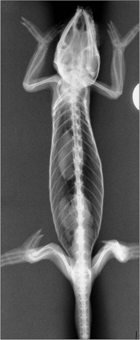
Figure 4.12 • Dorsoventral radiograph of Panther chameleon (Furcifer pardalis) showing zygodactyl feet.
Autotomy
Autotomy means “self-amputation” and is a mechanism to escape from predators. When the animal is attacked the brightly colored tail will break off and wriggle for a few minutes to distract the attacker, allowing the lizard to escape. It occurs in many lizards, such as the iguana, skink, and gecko species, where the tail is not essential for survival (Figs. 4.13 and 4.14). However, species like chameleons and monitors, which rely on their tail for climbing and defense, do not shed their tails. Similarly, the Marine iguana, which relies on its large rudder tail for swimming in the sea, lacks fracture planes.
Autotomy is created by a vertical fracture plane, containing no bone, passing through the body and part of the neural arch of each caudal vertebra (Bellairs 1998h; Bellairs & Bryant 1985; Evans 1986; Pough 1998b). This is a plate of cartilage or connective tissue that develops after ossification. These are not present in the cranial part of the tail so the cloaca and hemipenes are protected. In iguanas the fracture plane is replaced by bone during maturation, resulting in a more stable tail in adults.
CLINICAL NOTE
After autotomy the stump should never be stitched as the broken tail rapidly forms its own scab that is followed by growth of new epidermis within a week or two. Bleeding is minimal, owing to the action of sphincter muscles in the caudal arteries and valves in the veins. After about 2 weeks regeneration begins and a cylinder of cartilage is formed. This may become calcified, but as it has no individual tail vertebrae it is less flexible than the original model. It is innervated mainly by the last spinal nerves. It is finally covered by scales, which are often smaller and a different color from the original tail (Bellairs I998h; Bellairs & Bryant 1985; Pough 2002).
63
KEY POINTS
• Kinetic jaw for wide gape
• Large well-developed adductor (jaw-closing) muscles, so mind your fingers!
• Hind foot has first four metatarsal bones lying together while fifth metatarsal lies separated with backwardpointing hook
• Tail can self amputate (autotomy)
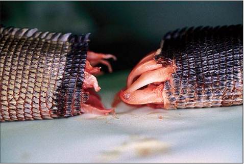
Figure 4.13 • Ends of tail post autotomy in a Green iguana (iguana iguana).
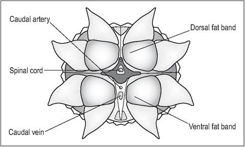
Figure 4.14 • Diagram of end on view of tail stump post autotomy.
GENERAL INTEREST
The Horned lizard (Phrynosoma cornutum) is a desert ground dweller that is found in the southern USA and Mexico and which can squirt blood from its eye when under attack. This is achieved by a pair of muscles, which restrict the blood outflow from the internal jugulars, causing an increase in blood pressure and leakage from the ocular venous sinuses. When threatened the lizard closes its eyes, which become swollen, and then shoots out a fine stream of blood from the eyelid margins (Barten 1996; Evans 1986; White 1976).