CARDIOVASCULAR SYSTEM
Snakes have a 3-chambered heart with a complete atrial separation and just one ventricle. It is long and slender and lies just cranioventral to the bifurcation of the trachea,
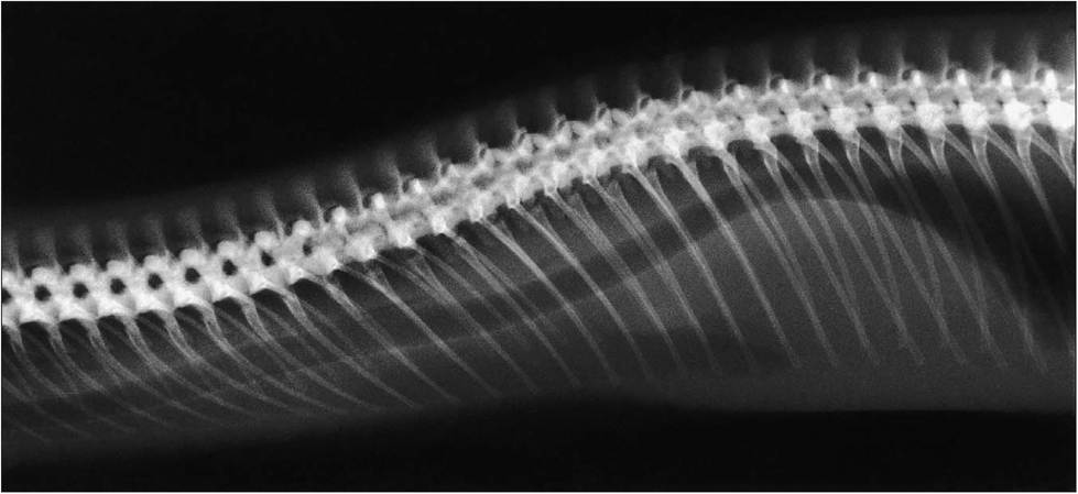
Figure 5.13 • Radiograph of vertebrae and ribs which extend the length of the body to the vent.
Post cloacally the vertebral processes are forked to protect the lymph hearts. Note the precardial fat pad.KEY POINTS
Anatomical adaptations for locomotion:
• Up to 400 flexible vertebrae
• Ribs that extend to midline from atlas to vent
• Large interconnecting muscle bundles
• Ventral gastropeges linked by pleated membranes
• Elastic dermis and loose skin
CLINICAL NOTE
Avoid the ventral approach when operating so as to avoid the ventral abdominal vein. When doing a celiotomy the incision is made between the second and third dorsal row of lateral scales. This also preserves the ventral scales for locomotion and keeps the wound from getting soiled by the substrate (Funk 1996).
about a third of the way down the body. The heart is fairly mobile as there is no diaphragm to hold it in place, and this allows prey items to pass by it.
Paired right and left aorta fuse caudal to the heart to form the dorsal aorta. A large ventral abdominal vein lies along the inner surface of the midline and so this must be avoided when making a celiotomy incision. A renal portal system is present, as in all reptiles.
The carotid arteries are asymmetrically placed. The more advanced snakes like the colubrids and vipers have only the left carotid artery, the right one being rudimentary.
As is the case with other reptiles, lymphatic vessels are prominent. Dilations of the lymph vessels (lymph hearts) are found on each side of the tail base where they are protected by modified, forked caudal vertebrae.
Venepuncture sites
(See Redrobe & MacDonald 1999.)
Ventral coccygeal vein - Insert the needle caudally to avoid the hemipenes in males.
Cardiac puncture - This is usually recommended in species over 300 g in weight. The needle is inserted into the ventricular apex and the syringe allowed to fill passively according to the cardiac cycle. There is little danger of leakage post sampling due to the slow heart rate and low blood pressure (Murray 2000).
Dorsal palatine vein - This can be easily visualized in medium to large sized snakes. It lies on the dorsal oral cavity, medial to the palatine row of teeth. Hematoma formation is common post sampling and saliva may contaminate blood samples (Murray 2000).
Jugular cutdown - This is possible on collapsed or anesthetized animals.