CARDIOVASCULAR SYSTEM
The heart rate can vary from 180 to 250 beats per minute. It is relatively small and the right atrioventricular valve has only 2 cusps, making the term “tricuspid” incorrect. The pulmonary artery is thickened and more muscular than in dogs and cats.
Rabbits have a simple conduction system and the sinoatrial node consists merely of a small group of cells generating impulses; this is why they were used in the first pacemaker experiments (Cruise & Nathan 1994).Unlike the dog, which has significant anastomoses between the internal and external jugular vein, the main vessel for return of blood from the head is the external jugular vein. Therefore, damage or ligation of this vein in rabbits means the eye is subject to proptosis. The same pattern also occurs with the external and internal carotid artery (Donnelly 1997).
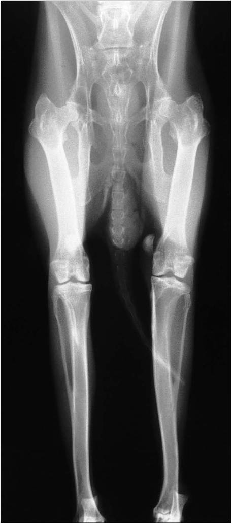
Figure 8.7 • This rabbit had a medial luxating patella of the left stifle which needed surgical correction. In rabbits there is a degree of fusion of the fibula and tibia.
Venepuncture sites
The blood volume of a rabbit is 55-70 ml/kg (Benson & Paul-Murphy 1999; Donnelly 1997). For blood sampling the best sites are the central auricular artery and jugular vein. Smaller samples (rabbit selects the most nutritious part of the plant, favoring young, succulent plants over mature, coarse growth (Cheeke 1987f). This browsing behavior (also seen in giraffes and deer) helps an animal of small body weight sustain its high metabolic rate (Cheeke 1987a, 1994). Rabbits ingest coarse fiber only to stimulate gut motility and, unlike horses (which carry fiber for up to three days) rapidly excrete it, thus obviating the need to carry vast quantities around (Brooks 1997; Harkness & Wagner 1995; Jenkins 2000).
| Table 8.1 Major differences in gastrointestinal tract between rabbit and ruminant (cow) | |
| Rabbit | Cow |
| Digest cellulose Cecal fermentation Main VFA is acetic acid Low gut retention time Bacteroides spp. microflora | Digest cellulose Rumen fermentation Main VFA is propionic acid Holds food for 4? longer than rabbit Lactobacillus spp. microflora |
VFA, volatile fatty acid
CLINICAL NOTE
Rabbits fed free choice will select concentrates (Cheeke 1987a; Harcourt-Brown 1996). This ability to select high protein and carbohydrate over fiber means that pet rabbits fed only a high concentrate diet may not get enough fiber to stimulate gut motility and will suffer consequently from intestinal stasis (Cheeke 1994).
The rabbit’s hindgut consists of a vast cecum, where food is fermented, and the proximal colon, which mechanically separates the high and low fiber particles. High fiber particles are eliminated fast while the nutrient-rich particles are sent back to the cecum to be made into cecotrophs. These high fiber particles, known as the “scratch factor” in the French rabbit industry are essential for the normal functioning of the rabbit gastrointestinal process (Brooks 1997).
Prehension of food
Rabbits have a blind spot directly in front of the mouth so cannot see food placed directly there. Instead it uses its sensitive prehensile lips and vibrissae for food discrimination and prehension (Jenkins 2000; Whitehouse & Grove 1968).
CLINICAL NOTE
Rabbits do not use their incisor teeth for prehending food, so in cases of severe incisor malocclusion it is possible to extract all incisor teeth. They will no longer be able to slice their food but this is rarely a problem for the pet rabbit (Fig. 8.16).
Oral cavity
The mouth aperture is small and designed for nibbling. The rabbit’s teeth are developed for the high fiber herbivorous diet. Wild rabbits have brown staining on the crowns of the teeth from feeding on natural grasses. The dental formula is 2/1,0/0,3/2, 3/3 = 28. Like all Lagomorphs, rabbits have three pairs of incisors: two upper and one lower (Crossley 2003; Vaughan 1986). The second upper incisors are rudimentary and called peg teeth and lie just behind the upper incisors.
At rest, the lower incisors should rest just behind the upper incisors (Fig. 8.12).There are no canines and the gap between the incisors and premolars is called the diastema (Fig. 8.11). The furred cheeks can be folded into this space, separating the incisor teeth from the oropharynx. The premolars and molars function as one unit and are often referred to as cheek teeth (Fig. 8.15). Branches of the lingual artery run close to the lower cheek teeth so must be avoided during dental surgery.
KEY POINTS
• Rabbits are highly efficient food convertors.
• Fiber is essential for gut motility.
• Browsing feeding means rabbits will selectively prefer high concentrates.
• Incisors and molars are continually growing. Malocclusion results secondary to an insufficiently fibrous and abrasive diet.
CLINICAL NOTE
Normal upper incisors have a vertical groove running along the length of the tooth and have smooth white enamel. If horizontal grooves are visible in the enamel it can be a sign of dental disease due to poor diet (Harcourt-Brown 1996, 1997).
CLINICAL NOTE
Mandibular prognathism means an abnormally long jaw relative to the length of the maxilla. It is inherited as an autosomal recessive trait and leads initially to edge-to-edge apposition and blunting of the chisel edges. Later, the lower incisors protrude rostral to the upper incisors (Fig. 8.17). The mandible is united by a strong fibrous symphysis and is narrower than the maxilla (Fig. 8.13 and 8.14). Both incisor and molar teeth are rootless (aradicular) and constantly growing at a rate of approximately 2 mm per week. The incisors help to slice the food, which is then chewed to a bolus by the action of the cheek teeth, which along with the tongue move the bolus caudally so that all teeth get equal chewing action (Crossley 2003).
Mastication
The flexible temporomandibular joint allows the jaw to open and close and also move rostrally, caudally, and laterally (Fig. 8.11). The cheek teeth arcades function as one unit and grind the long stem fibers into small food boluses, which
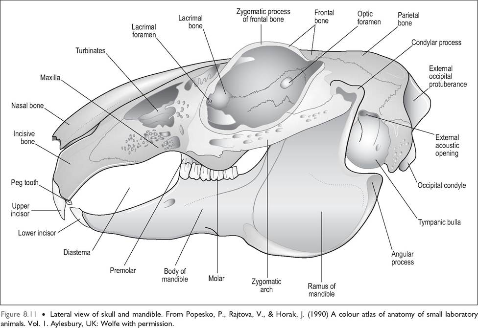
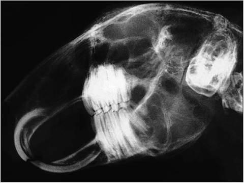
Figure 8.12 • Lateral radiograph of skull and mandible.
can then be swallowed. The jaws appose only one side at a time and rotate laterally at a rate of 120 times a minute. The tongue is utilized to make sure all food is thoroughly masticated. Cecotrophs are not chewed but swallowed intact (Brewer & Cruise 1994).
Tongue
The tongue is very long and has an elevated region caudally called the lingual torus. Numerous papillae along its length give it a roughened appearance. Four types are present: vallate, foliate, fungiform, and filiform; all but the last contain taste buds (Crossley 2003). Unlike the rat, the rabbits has paired tonsils.
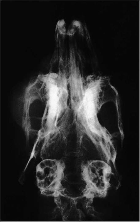
Figure 8.14 • Dorsal radiograph of skull (slightly rotated).
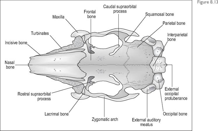
Dorsal view of skull.
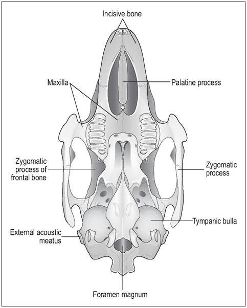
Figure 8.15 • Ventral view of skull. From Popesko, P., Rajtova, V., & Horak, J. (1990) A colour atlas of anatomy of small laboratory animals. Vol. 1. Aylesbury, UK: Wolfe with permission.
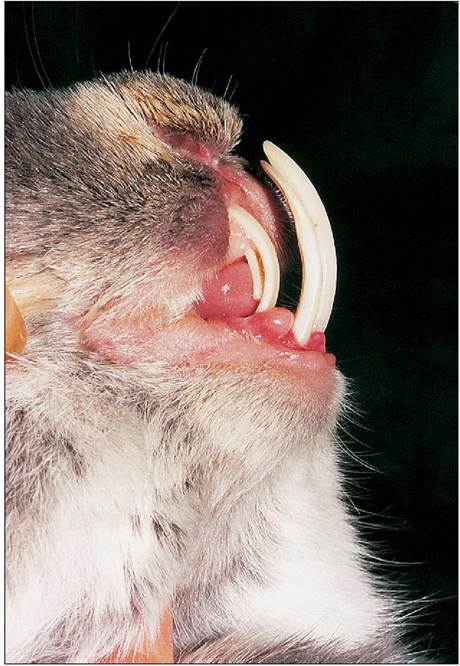
Figure 8.16 • Close up of rabbit with severe incisor malocclusion showing upper and lower incisors and peg teeth. (Photo by Claire Nuttall)
Salivary glands
There are four pairs of salivary glands: the parotid, zygomatic, mandibular, and sublingual (Jenkins 2000). The sublingual is a minor gland as the major sublingual is not present. The zygomatic gland lies just below the lacrimal gland in the anteroventral angle of the orbit (Cruise & Nathan 1994). Amylase is secreted by the glands in response to food entering the mouth (Brewer & Cruise 1994).
Esophagus
The esophagus has three layers of striated muscle, which, unlike in the dog and humans, extend all the way to the cardia of the stomach. There are no mucous glands in the esophagus.
The cardia has a well-developed sphincter and is positioned so that the rabbit cannot vomit (Brewer & Cruise 1994; Cruise & Nathan 1994)(Fig. 8.18).Abdominal cavity
Rabbit muscles are pale red in comparison to the darker red of cats and dogs (Okerman 1994). There is scant subcutaneous tissue on the midline. The linea alba is thin and lies in very close proximity to the cecum and bladder.
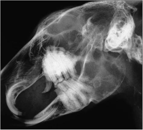
Figure 8.17 • Lateral radiograph of skull of dwarf rabbit with incisor and molar malocclusion secondary to a combination of congenital mandibular prognathism and poor diet since birth.
CLINICAL NOTE
When performing abdominal surgery, elevate the linea alba well to avoid incising the viscera underneath. The tendon is so thin that stitching the midline means stitches in the rectus abdominis muscle. On opening into the cavity, the main organs (cecum and colon) run obliquely from the right liver lobes, caudally to the left side of the bladder (Fig. 8.21).
Stomach
The stomach is ‘J'-shaped, thin walled and lies on the left side. The cardia is lined by non-glandular stratified squamous epithelium. The fundus is glandular and its parietal cells secrete acid and intrinsic factor; the chief cells secrete pepsinogen. The pylorus is heavily muscled with a well-developed sphincter (Cruise & Nathan 1994). The stomach normally contains a mixture of food, fur, and fluid, even 24 hours post feeding. The pH 1-2 of the adult rabbit makes the stomach and small intestine almost sterile (Jenkins 2000)(Fig. 8.18).
CLINICAL NOTE
Gas distension after death due to autolysis often ruptures the thin stomach wall, so this is a common and normal postmortem finding. The presence of hair in the stomach is normal and due to rabbit grooming behavior. However, large hair impactions (hairballs or trichobezoars) are indicative of dehydration and lack of gastric motility (Brooks 1997; Donnelly 1997).
Neonatal stomach
The neonate stomach has a pH 5.0-6.5 (Brewer & Cruise 1994; Harkness & Wagner 1995; Jenkins 2000) and a stomach full of milk curd. This would make an ideal breeding ground for bacteria but for the fact that, in the first 3 weeks of life, it is acidified by the production of milk oil. This consists of octanoic and decanoic fatty acids produced by the enzymatic reaction of the suckling rabbit's digestive enzymes with the doe's milk (Harkness 1990; Harkness & Wagner 1995). Hand-reared rabbits lack this protective antimicrobial factor, making them very susceptible to infections. From about 2 weeks of age young rabbits also begin to acquire some gut flora by eating the doe's cecotrophs. By the time milk oil production ceases at 4-6 weeks some organisms will have managed to colonize the cecum to produce the hindgut fermentation. At weaning the pH drops to adult pH 1-2, which keeps the stomach relatively microbe free (Brooks 1997; Cheeke 1987a).
CLINICAL NOTE
Weaning is a critical time in the rabbit’s life. The protective effect of milk oil has waned and the pH has not yet reached the adult pH 1-2. If the gut is not colonized by healthy bacteria, coliforms and clostridia can proliferate causing rapid enterotoxemia. This is easily precipitated by a low fiber, high starch diet (Brooks 1997).
Small intestine
The small intestine is relatively short and comprises only 12% of the gastrointestinal volume. This is the site of digestion and absorption of sugars and protein from food
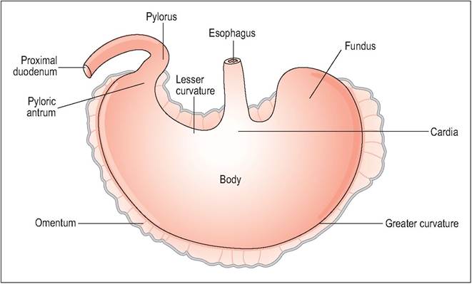
Figure 8.18 • External view of rabbit stomach showing location of cardia in the center of lesser curvature. This means that rabbits cannot vomit so need not be fasted prior to anesthesia. From Popesko, P., Rajtova, V., & Horak, J. (1990) A colour atlas of anatomy of small laboratory animals. Vol. 1. Aylesbury, UK: Wolfe with permission.
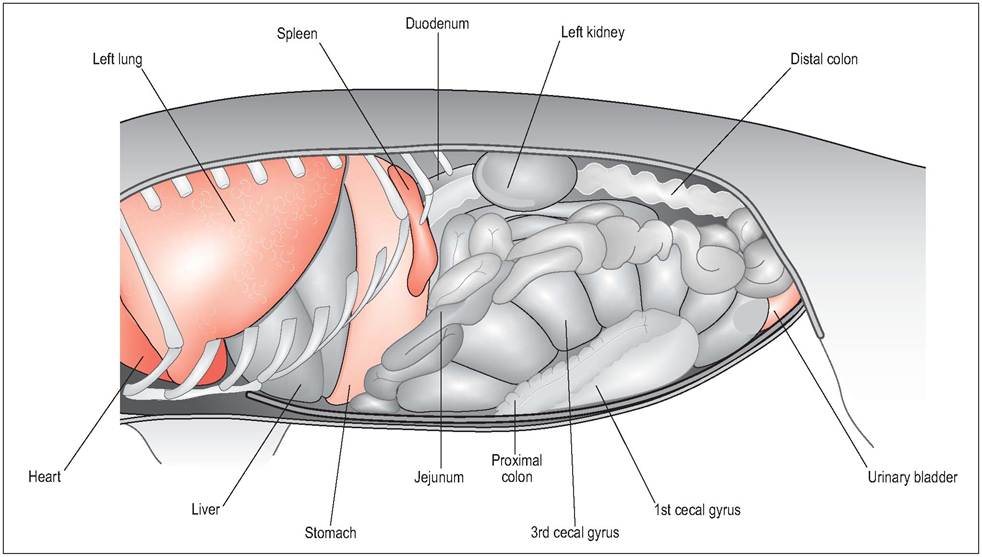
Figure 8.19 • Left lateral view of abdominal viscera (with some ribs removed). From Popesko, P., Rajtova, V., & Horak, J. (1990) A colour atlas of anatomy of small laboratory animals. Vol. 1. Aylesbury, UK: Wolfe with permission.
items, as well as of vitamins, proteins, and fatty acids from the cecotrophs (Cheeke 1987a). Moitilin is secreted by the endocrine cells of the duodenum and jejunum and helps to stimulate gastrointestinal motility in the small intestine, colon, and rectum (but not in the cecum). High carbohydrate diets inhibit its secretion and lead to gut stasis problems; fats, however, stimulate moitilin release (Brewer & Cruise 1994).
The duodenum lies at an acute angle to the liver and can be compressed by it. Unlike in most mammals, the bile duct and pancreatic duct enter the duodenum at widely separated points: the bile duct near the beginning and the pancreatic duct at the end of the duodenum (Cruise & Nathan 1994).
CLINICAL NOTE
Rabbits are unable to vomit and the duodenum becomes easily compressed by the liver. Stomach distension and lifethreatening bloat can occur with hepatomegaly or if the stomach becomes impacted by hair.
The jejunum is slightly less thick and vascular than the duodenum. Aggregates of lymphoid tissue (Peyer patches) in the lamina propria become most prominent towards the ileum. The terminal ileum enlarges into a dilation called the sacculus rotundus, which is unique to the rabbit (Fig. 8.22). This lies in the left caudal abdominal quadrant and is often called the cecal tonsil; it has many lymph follicles that give it a honeycomb appearance. This opens into the ampulla coli at the junction between the ileum, colon, and cecum. A weak valve, the ileocecal valve, allows the chyme to pass one way into the cecum (Cruise & Nathan 1994).
Hindgut
The hindgut is well developed and consists of the cecum and colon (Figs. 8.19 and 8.20).
Cecum
The rabbit cecum is the largest of all animals, relative to size, with 10 times the capacity of the stomach and containing 40% of the intestinal content (Cruise & Nathan 1994; Jenkins 2000). It is very thin walled and coils on itself in three gyral folds before ending in a blind-ended, thick-walled tube called the vermiform appendix. The appendix is rich in lymphoid tissue and also secretes bicarbonate to buffer the cecal acids and water to form the semi-fluid cecal paste (Harkness & Wagner 1995). Unlike many other herbivores, the main cecal microorganisms in the rabbit are not lactobacillus but Bacteroides spp., plus ciliated protozoa, yeasts, and small numbers of Escherichia coli and clostridia (Cheeke 1987a; Harkness & Wagner 1995).
CLINICAL NOTE
The cecal wall is very thin and can tear easily so it is important to avoid handling, or even touching it, during surgery if at all possible.
The cecum acts like a large fermentation vat where the microbial flora break down cellulose and proteins into volatile fatty acids (VFAs) which are then directly absorbed across the cecal epithelium into the bloodstream. Its contents are semi-fluid and have an alkaline pH in the morning and acid pH in the mid afternoon. Changes in pH cause “trans- faunation”, which is a change in the type of microorganisms present (Brewer & Cruise 1994).
Unlike ruminants, the predominant VFA in the rabbit is acetate, regardless of diet, followed by butyrate and propionate. This is caused by the dominance of Bacteroides spp. instead of lactobacillus in the cecum (Carabano & Piquer 1998; Cheeke 1987d) (Table 8.1).
Cecotrophy
Coprophagy is the consumption of feces; cecotrophy refers to the consumption of cecal pellets. Cecotrophy starts between 2 and 3 weeks of age when kits start to eat solids (Carabano & Piquer 1998). They start by ingesting maternal cecotrophs first. Cecotrophy is essential for rabbit health and the lack of cecotrophy leads to a lower level of nutrients and reduced availability of protein and B and K vitamins (Eden 1940).
Cecotrophs (or night stools) are formed in the proximal colon and cecum. While the high fiber pellets (>0.5 mm in size) do not enter the cecum, but are excreted rapidly, the fine fiber particles and fluid remain in the cecum to form high nutrient particles. These become coated with mucus
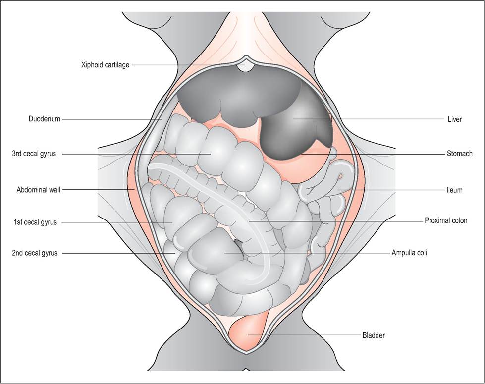
Figure 8.20 • Diagram showing ventral view of abdominal viscera.
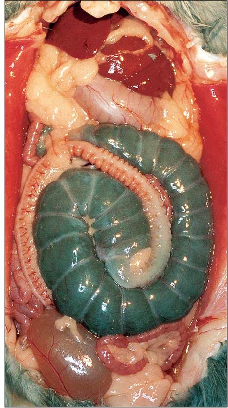
Figure 8.21 • Ventral view of abdominal viscera.
produced by the goblet cells of the colon before passing out through the anus as grape-like clusters. They are ingested directly from the anus by anal reflex and are swallowed whole without mastication. When they reach the fundus of the stomach, where further fermentation takes place over 3-6 hours, the mucus coating protects them from digestion (Cheeke 1987a).
of the gut, and reduces the amount of cecotrophs produced. Indigestible fibers like cellulose and lignin (crude fiber digestibility of 15%) are the best way of preventing enteritis as they stimulate most hindgut motility. Nonlignified fiber like beet pulp (crude fiber digestibility of 60%) are less effective (Cheeke 1994).
A low protein diet encourages the rabbit to eat more cecotrophs in order to alleviate the deficiency whereas a diet high in protein and low in fiber reduces their consumption (Cheeke 1987b).
High carbohydrate diets produce two-fold problems. Excess glucose provides a medium for the bacteria like Clostridium Spiroforme and E. coli to colonize. These diets also produce excess VFAs in the cecum, leading to a drop in pH, which in turn inhibits normal flora and allows pathogens to proliferate (Cheeke 1987g, 1994). High starch foods like maize (corn) is the least satisfactory; oats and barley are better as they are higher in fiber while being lower in starch (Cheeke 1987d, 1994).
Fats can be used as source of energy without causing cecal hyperfermentation. Full-fat soybeans and oilseeds are good sources and vegetable oil is better than animal fat (Cheeke 1994).
Colon
Although anatomically the colon has an ascending, transverse, and descending portion, functionally it is divided into a proximal part (approx. 50 cm) and a longer distal part (approx. 90 cm) (Fig. 8.22). The proximal colon has three parts: three longitudinal muscular bands called taenia creating three haustra (sacculations), a single taenia/haustra and the fusus coli. The distal colon has no sacculations (Carabano & Piquer 1998; Ehrlein et al. 1983; Ruckebusch & Hornicke 1997).
The fusus coli is unique to lagomorphs. It is a 5-8 cm area of thickened circular muscle lined by thick mucosa (Cruise & Nathan 1994). It is heavily supplied with ganglion cells and is under the influence of aldosterone and prostaglandins. It serves as a pacemaker, regulating the passage of ingesta into the distal colon. It controls three types of colonic motility: segmental, peristaltic, and haustral and it is these differing form of contractions which produce the hard and soft feces (Ehrlein et al. 1983; Ruckebusch & Hornicke 1997).
Circadian rhythm
Although wild rabbits produce cecotrophs during the day when sleeping in their burrow, pet rabbits tend to produce hard pellets 4 hours post feeding and cecotrophs 8 hours post feeding, which tends to be at night.
Effect of diet on cecotrophy
High fiber diets are essential for cecotrophy. Low fiber diets increase cecal retention time, leading to hypomotility
CLINICAL NOTE
Colonic motility and cecotrophy are regulated by the autonomic nervous system and aldosterone. This means that any kind of stress, like surgery or diet change, increases adrenaline (epinephrine), which can inhibit gastrointestinal motility and lead to cecal stasis and abnormal cecotrophs (Cheeke I987g; Lebas et al. I997a).
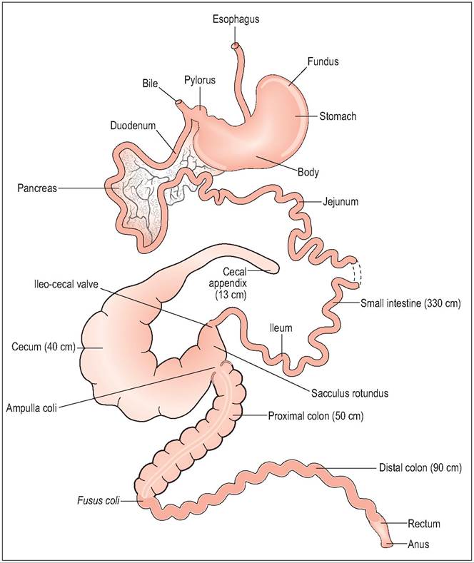
Figure 8.22 • Diagram of digestive tract of the rabbit.
CLINICAL NOTE
Production of hard feces
Segmental and haustral contractions in the proximal colon (particularly of the single haustra) mechanically separates the ingesta into solid indigestible particles and liquid contents. The large, solid pellets pass down in the middle of the lumen, further water is absorbed and they are excreted as hard dry pellets. The liquid fraction and smaller particles move to the periphery into the haustrae where they are returned by antiperistalsis into the cecum for further fermentation (Cheeke 1987a; Ehrlein et al. 1983; Ruckebusch & Hornicke 1997).
Production of soft feces (cecotrophs)
Segmental and haustral contractions are reduced and the cecum contracts to expel a soft paste into the proximal colon. Motility is decreased in the proximal colon under prostaglandin influence but enhanced in the distal colon so that the digesta passes rapidly down with no separation and no water absorption (Ehrlein et al. 1983; Ruckebusch & Hornicke 1997).
Athough vomiting is not possible and diarrhea is rare in adult rabbits, decreased water and electrolyte absorption from the colon with intestinal hypomotility leads rapidly to dehydration. Consequently, fluid therapy is essential for rabbits with gastrointestinal disease (Cheeke 1987g, 1994).
KEY POINTS
• Weanling rabbits become vulnerable as the protective effect of mothers “milk oil” wanes.
• The fusus coli controls separation of hard and soft feces.
• Indigestible fiber (cellulose and lignin) is essential to drive the gut.
• High protein and high starch foods lead to gastrointestinal tract motility problems.
• High fiber and fats increase gastrointestinal tract motility.
• Bacteroides spp. are the main bacteria of the hindgut.
• Stress, via aldosterone, inhibits gastrointestinal motility, leading to intestinal stasis.
Liver
CLINICAL NOTE
The liver has four lobes with a deep cleft dividing it into right and left lobes. Each lobe is then subdivided into medial and lateral. The right lobe has two further subdivisions, with the quadrate lobe in the midline and the caudate lobe near the right kidney. This small lobe has a narrow attachment to the hilar region of the liver and could be a site of liver torsion.
The gall bladder is located very deep in the right anterior lobule. The hepatic ducts unite to form the common bile duct, which receives the cystic duct from the gall bladder and enters the proximal part of the duodenum (Cruise & Nathan 1994).
Rabbits secrete about seven times as much bile as a dog of similar weight. (A 2 kg rabbit secretes 250 ml bile daily) (Brewer & Cruise 1994). They resemble birds and reptiles in that they secrete mainly biliverdin (63%) rather than bilirubin. This is due to low levels of bilirubin reductase, which reduces biliverdin to bilirubin (Cheeke 1987a; Jenkins 2000).
CLINICAL NOTE
A cardinal sign of dental disease can be polydipsia, secondary to painful teeth causing anorexia.
Kidney
The right kidney lies cranial to the left (Fig. 8.23). The rabbit kidney is quite primitive in comparison to that in other mammals. However, it has been extensively researched as its kidney tubules can be easily removed with the basement membrane intact, facilitating renal research. The kidneys are unipapillate meaning that one papilla and calyx enter the ureter (Brewer & Cruise 1994; Cruise & Nathan 1994).
A feature of the rabbit kidney is that, like neonatal mammals, not all glomeruli are active at one time. This means that a well hydrated rabbit can activate dormant glomeruli and increase diuresis without having to increase renal plasma flow and glomerular filtration rate (Brewer & Cruise 1994; Cruise & Nathan 1994).
Reabsorption of bicarbonate from the renal tubules is not as efficient as in other mammals due to the lack of carbonic anhydrase. This enzyme catalyzes the conversion of carbon dioxide to bicarbonate, and vice versa, acidifying luminal fluid in the collecting ducts. As rabbits also produce high levels of bicarbonate from bacterial fermentation this means they easily get a surplus of bicarbonate and metabolic alkalosis (Brewer & Cruise 1994). Rabbits consequently excrete a much more alkaline urine than other animals like rats (Cheeke 1987e).
Anorexia in rabbits can rapidly lead to fatal hepatic lipidosis. This is caused by prolonged hypoglycemia inducing lipolysis of fat stores into fatty acids. When the body cannot metabolize these excess free fatty acids they accumulate in hepatocytes of the liver, literally turning it into lard. Obese rabbits will already have some degree of fatty liver so even mild anorexia can cause life-threatening hepatic lipidosis.
Pancreas
This is a diffuse, irregular mass lying in the duodenum and is relatively small. The pancreatic duct enters the distal duodenum some 35-40 cm from the bile duct (Cruise & Nathan 1994).