CARDIOVASCULAR SYSTEM
Heart
It has been said that the unpaired innominate artery (brachiocephalic) at the base of the neck aids the ferret's agility (Willis & Barrow 1971) but there are still two common carotid arteries in the neck and not a single one as implied.
In the ferret the heart can be compromised with the occurrence of dilated cardiomyopathy (DCM) and hypertropic cardiomyopathy (HCM) in both sexes (Fig. 12.5). DCM progresses slowly over time whereas HCM initiates sudden death by left ventricle hypertrophy. The ferret heart can also become dysfunctional due to the presence of Dirofilaria immitis adults in the chambers, as with other carnivores.
Being a small mammal the ferret heart rate is usually between 200 and 400 beats per min. The cardiovascular/ respiratory and arterial blood pressure standards are given in Table 12.1.
It is difficult to assess cardiac performance in the ferret using techniques such as systemic arterial blood pressure, central venous pressure, right ventricular pressure or pulmonary capillary wedge pressure. Estimations of cardiovascular performance, and more specifically, cardiac output, are usually limited to monitoring pulse strength and urine output. Unfortunately, the pulse is not reliably palpable in ferrets so urine output is therefore the best indicator of cardiac output (Lucas 2000).
The 4-chambered heart consists of left and right auricles and left and right ventricles, as is usual in mammals. The heart muscle is typical and, like the lungs, the ferret heart has been used in research (Whary & Andrews 1998). It lies between the 6th and 8th ribs and is obliquely placed in the thoracic cavity with the apex to the left side (Fig. 12.4). For auscultation purposes it lies more caudal in the chest than first imagined. The heart ligament joining it to the sternum will lose its fatty coat in cases of heart disease and, on radiology, if the heart is actually resting on the sternum, it can be a sign of early cardiac enlargement and disease according to Brown (1997).
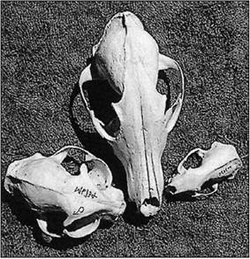
Figure 12.2 • Comparative skulls of three pet carnivores: ferret, dog, and cat.
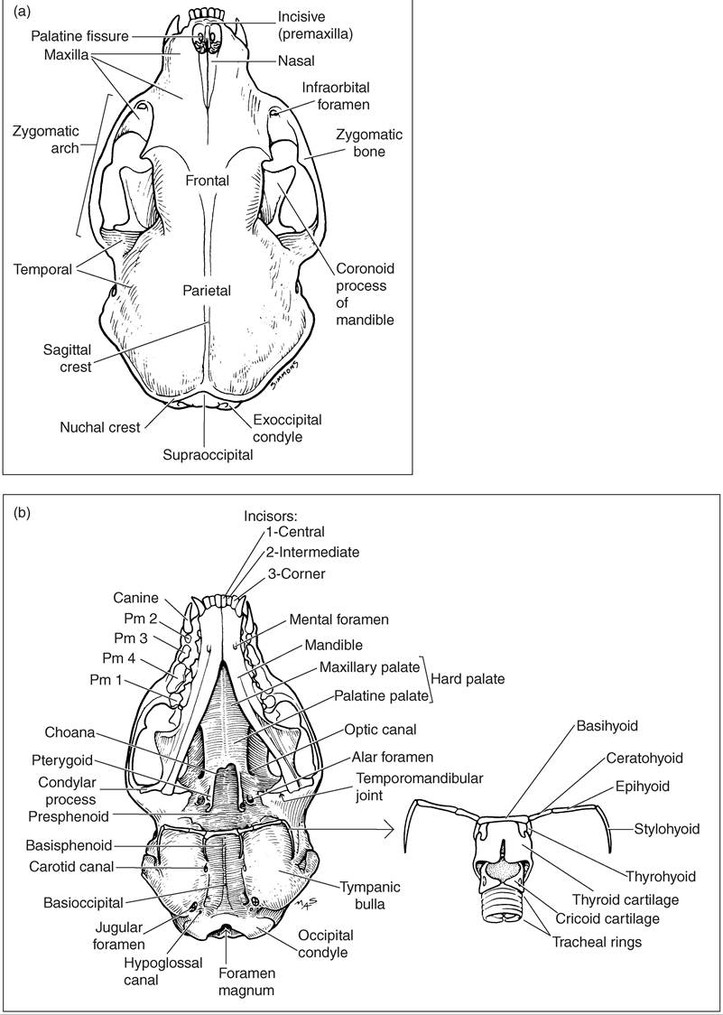
Figure 12.3 • (a) The ferret skull dorsal view, (b) The ferret skull ventral view. (Courtesy of Howard Evans.)
Venepuncture
Hematological and serum chemistry require venous blood sampling, using cephalic and jugular veins (Figs. 12.6 and 12.7). If you refer to Fig. 12.10, this also shows the external jugular vein, one of the main routes for bleeding or giving blood. It lies quite lateral on the neck and can be difficult to palpate in the hob due to its thickened neck. There is a laboratory technique for tail artery sampling, which is useful for periodic sampling of numerous ferrets (Curl & Curl 1985).
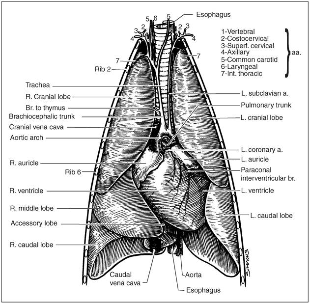
Figure 12.4 • Ferret heart and lungs (ventral view). (Courtesy of Howard Evans.)
The healthy ferret should have a packed cell volume (PCV) greater than 20%. For a sick ferret, with PCV below 15%, a blood transfusion is advised. Blood typing is not necessary in ferrets for blood transfusions (Manning & Bell 1990). Samples of 6, 9 or 12 ml of blood can be taken from jills, small or large hobs, respectively (Lucas 2000). Note that, when sedated, an average 1.5-2.0 kg hob can give 15-20 ml of blood and the average 0.75-1.00 kg jill, 10-12 ml (Jenkins & Brown 1993).