RESPIRATORY SYSTEM
Lungs
The lungs are relatively long in the tube-like space of the chest in such a sleek animal as the ferret. They give lateral and dorsal cover to the heart and are divided into cranial, middle, caudal, and accessory lobes on the right side, and cranial and caudal on the left (Fig.
12.4). The thoracic inlet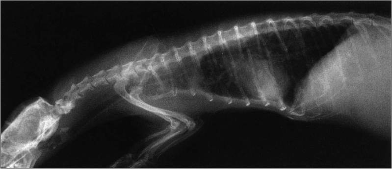
Figure 12.5 • Radiograph of ferret showing chest structure. This ferret has cardiomyopathy.
Adapted from Fox, J. G. (1988). Biology and diseases of the ferret, 2nd edn. p. 184, with permission
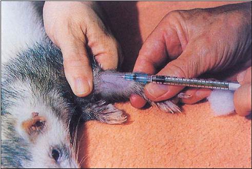
Figure 12.6 • Taking blood from the cephalic vein. Photo courtesy of John Tingay.
is narrower compared to other carnivores which have more bulk, such as dogs, and contains anterior mediastinal lymph nodes and passage of the trachea, esophagus, and major blood vessels. Any abnormality of even one organ at this point can cause serious interference with chest function. Major neoplastic conditions can arise in young and old ferrets, such as lymphoma and fibrosarcoma.
The ferret lungs have a large volume in relation to body weight, with the total lung capacity exceeding a predicted value by 297%, hence their value as experimental animals for research into human conditions. Interestingly, ferret lung structure contains excess submucosal glands in the bronchial wall and extra terminal bronchioles, making them anatomically like human lungs (Whary & Andrews 1998). Ferrets can be infected with human influenza virus as well as canine distemper.
CLINICAL NOTE
During operations care must be taken not to compress the ferret's chest as they rely more on diaphragm movement for ventilation under anesthesia than on costal movement. If a ferret stops breathing, cardiopulmonary resuscitation can be instigated by holding it by the legs and moving it side to side to stimulate diaphragmatic breathing.