Classification of Joints
Based on their structure and the material that unites them, joints may be classified as fibrous, cartilaginous, or synovial. These classifications can be combined as follows.
Fibrous Joints
Fibrous joints have no joint cavity.
The bones are united by fibrous tissue (Fig. 6-1).Syndesmosis refers to a joint united by fibrous tissue that permits only slight movement. The normal union of the shafts of the splint bones and cannon bone of the horse is an example of syndesmosis.
A suture is the particular fibrous joint between bones of the skull. Sutures often completely ossify in maturity.
A gomphosis is the specialized articulation of teeth in their alveoli (sockets) in the
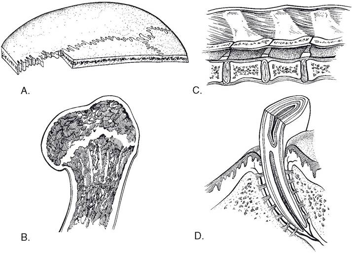
Figure 6-1. Types of non-synovial joints. A) Suture. B) Synchondrosis (growth plate). C) Symphysis (an intervertebral disk). D) Gomphosis.
mandible and maxilla. The collagenous tissues and fibroblasts that join the tooth to the socket constitute the periodontium.
Cartilaginous Joints
The bones of a Cartilaginousjoint are united by cartilage, with no intervening joint cavity.
A synchondrosis is an immovable joint in which the uniting medium is hyaline cartilage. The union of the diaphysis and epiphysis of an immature bone (its physis or growth plate) is an example of synchondrosis (Fig. 6-1).
Symphyses (fibrocartilaginous joints) are united by flattened disks of fibrocartilage as found between adjacent pelvic bones and between the bodies of adjacent vertebrae and sternebrae.
The fibrous or cartilaginous tissues separating adjacent bones in syndesmoses, synchondroses, and symphyses can be replaced by bone as a result of either aging or degenerative processes. When this occurs, the joint is sometimes called a synostosis.
Synovial Joints
Most synovial (formerly called diarthrodial) joints have similar general structure, which includes articular surfaces, articular cartilages, articular cavity, joint capsule, and ligaments (Fig.
6-2).The articular surfaces are specialized layers of compact bone on the surfaces that articulate with other bones.
The articular cartilage is a layer of hyaline cartilage covering the articular surface.
The articular cavity is a space between the adjacent bones of the joint surrounded by the joint capsule. Because the space is normally very small and has within it only a very small amount of lubricating fluid, it is called a potential space. inflammation can expand the space with accumulation of fluid, a condition called joint effusion.
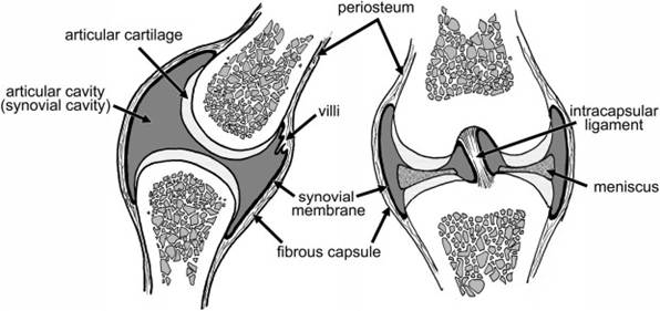
Figure 6-2. Synovial joints. In these figures, the heavy black line represents the synovial membrane. Note that it does not cover the articular cartilage or menisci, although it does surround the intracapsular ligaments. The fibrous capsule is continuous at its attachments with the periosteum of the bones.
The joint capsule consists of two layers. The deeper layer is the synovial membrane, a delicate layer of specialized connective tissue extending from the edges of the articular cartilages of the adjacent bones but not covering the articular cartilage. This membrane secretes the synovial fluid (synovia), which lubricates the normal joint. The synovial membrane’s surface area may be increased by folds (plicae synoviales), which may contain fat pads, that project into the joint cavity. Villi (villi synoviales), fingerlike projections, may also project into the joint cavity.
The superficial layer of the joint capsule is the fibrous capsule, a heavier fibrous sleeve adjacent to the synovial membrane. This fibrous layer may be thickened in certain areas to form the extracapsular (or periarticular) ligaments that connect adjacent bones and help stabilize the joint.
Ligaments, in relation to the musculoskeletal system, are connective tissue bands that extend from bone to bone. (Folds of serous membrane as seen in the thoracic, abdominal, and pelvic cavities are also called ligaments and are described with the appropriate organs in other chapters.) Tendons are connective tissue bands that connect muscle to bone.
They are described in Chapter 7.Intracapsular (intra-articular) ligaments are found within joints and are surrounded by the synovial membrane. The cruciate ligaments of the stifle are intracapsular ligaments.
Extracapsular (periarticular) ligaments are external to the joint capsule; they include collateral, dorsal, palmar, plantar, and annular ligaments. Collateral ligaments lie on the medial and lateral aspects of a joint. Dorsal and palmar (or plantar) ligaments lie in front of and behind the joint. Annular ligaments surround the joint, and their fibers generally circle the joint to strengthen and protect the capsule.
Menisci (fibrocartilage disks) are interposed between surfaces of some joints, where they contribute to the congruency of the articular cartilages and probably play a role in complex joint movements. Menisci are truly intracapsu- lar in that they are not covered by synovial membrane. Prominent menisci are found in the stifle and the temporomandibular joint.
Other Synovial Structures
Synovial membrane is also associated with two other structures, discussed more completely in Chapter 7. A bursa is a small, fluid-filled sac lined with synovial membrane. Bursae act as cushions and are generally found where tendons cross over a bony prominence.
Tendons may also be protected from bony prominences by a synovial sheath, a synovial membrane-lined tube that wraps around the tendon’s circumference. Synovial sheaths are particularly noteworthy in the distal limbs, where long tendons pass over joints. Inflammation of a synovial sheath and its tendon, or tenosynovitis, may follow trauma or penetrating injury and can result in a very obvious and painful distension of the sheath. Tendons and synovial sheaths are discussed more completely in Chapter 7.