» Clinical Considerations
Muscles of the epaxial and hypaxial divisions must often be separated and detached when access to the vertebral column is necessary. In the neck the ventral approach is most often chosen, although a dorsal approach is also possible.
In the lumbar region the dorsal approach is preferred.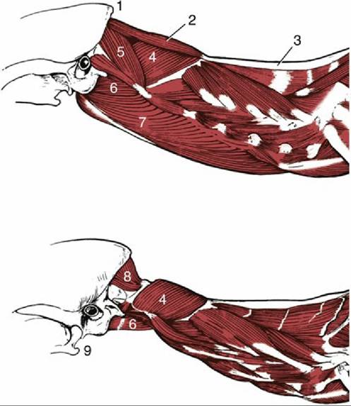
FIG. 12.12 Muscles associated with the canine atlanto-occipital and atlantoaxial joints, lateral view. 1, External occipital protuberance; 2, rectus capitis dorsalis major muscle (m.); 3, nuchal ligament; 4, obliquus capitis caudalis m.; 5, obliquus capitis cranialis m.; 6, rectus capitis ventralis m.; 7, longus capitis m.; 8, rectus capitis dorsalis minor m.; 9, angular process of mandible.
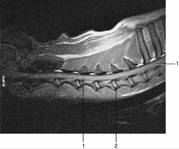
FIG. 12.13 Midsagittal section of cervical region of dog, T2-weighted magnetic resonance image. 1, Spinal cord; 2, nucleus pulposus.
The ventral approach to the cervical vertebrae (Fig. 12.13) is indicated for disk fenestration in cases of herniation or for the treatment of atlantoaxial instability. The trachea is exposed through a ventral midline incision, midway between the sternomastoid and sternohyoid muscles. Reflection of the trachea to the left protects the esophagus and exposes the paired longus colli muscles, which can be separated longitudinally.
The dorsal approach to the cervical vertebrae is indicated for vertebral fractures. This approach includes exposure of the biventer cervicis and the rectus capitis dorsalis major cranially, and the nuchal ligament, the spinalis et semispinalis cervicis, and multifidus cervicis muscles more caudally. The vertebral artery lies in the rectus capitis dorsalis major, ventrolateral to the synovial joint C1/C2 and must be avoided as the dissection is continued laterally.
The dorsal approach to the caudal cervical and cranial thoracic vertebrae for dorsal laminectomy (removal of part of the vertebral arch) and fracture repair first exposes the aponeuroses of the trapezius cranially and the rhomboid caudally. Then the subscapulares, splenius, and serratus dorsalis are exposed by lateral retraction of the trapezius and rhomboid muscles and the scapula. Finally, the semispinalis capitis and longissimus cervicis, the nuchal ligament, and the dorsal spines of the vertebrae are exposed by lateral retraction of the splenius and serratus dorsalis. The deep cervical artery passes through the semispinalis capitis.
A dorsal approach to the thoracolumbar vertebrae is indicated for dorsal laminectomy and thoracolumbar fractures. Lateral retraction of the lumbar fascia exposes the longissimus lumborum and multifidi caudally and the spinalis et semispinalis thoracis cranially. The multifidus, interspinalis, and rotatores longi are elevated from the spinous processes and vertebral arches. The dorsal branch of each spinal nerve emerges just cranial and ventral to the insertions of the longissimus on the accessory processes.
Comprehension Check
Use a cadaver to practice ventral and dorsal approaches to the cervical vertebrae of the dog.