» Conformation and Surface Anatomy
The shape of the thorax differs considerably among different breeds, as is well illustrated by the deep, laterally compressed thorax of the Greyhound (Fig. 13.1) and the broad, barrel-shaped one of the Pug (Fig.
13.2). These differences are reflected in the form of the ribs, which are long and relatively straight in the Greyhound, and shorter and strongly curved in the Pug. In cats, corresponding but less pronounced variation distinguishes the Oriental breeds from the Persian.The small size of the cranial part of the bony thorax and thus of the thoracic inlet is masked by the enclosure of the upper parts of the forelimbs within the skin of the trunk (Fig. 13.3) and by the height of the first few thoracic spinous processes (Fig. 13.4). The dorsal contours of the neck and thorax generally meet without a noticeable elevation at the withers. The skin is loosely attached here, making it a suitable site for the subcutaneous infusion of large volumes of fluid when necessary. The tips of the thoracic spinous processes are individually palpable, together with the spine and the cranial and caudal angles of the scapula to each side. In the standing dog, the cranial and caudal angles are opposite the spinous processes of the first thoracic vertebra and the bodies of the fourth and fifth thoracic vertebrae, respectively. The shoulder joint is located opposite the ventral end of the first rib, and the point of the shoulder is slightly behind the level of the manubrium of the sternum. The gently curved sternum rises between the forelimbs to the thoracic inlet, bringing the easily palpated manubrium a few centimeters cranial to the first pair of ribs. The olecranon projects on the thoracic wall immediately below the ventral end of the fifth intercostal space. However, breed and individual variations in the preceding features are common (Figs.
13.4 and 13.5).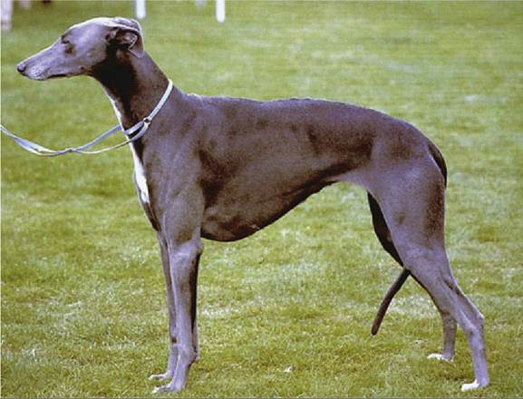
FIG. 13.1 Deep and laterally compressed thorax of the Greyhound.
The epaxial muscles provide a thick covering to the thoracic vertebrae and the dorsal parts of the ribs. The triceps muscles occupy the angle between the scapula and humerus, making it difficult to distinguish the caudal border of the scapula. Medial to the triceps and behind the limb, the lateral parts of the ribs are more thinly covered by the serratus ventralis, latissimus dorsi, scalenus, and obliquus abdominis externus muscles; one can feel the ribs through them (Fig. 13.6). The ventral surface of the thorax is covered by pectoral muscles. The axilla is deep and permits palpation of the first five ribs and the axillary and accessory axillary lymph nodes, especially when they are enlarged. The most extensive exposure of the chest is obtained when the limb is drawn forward.
The thorax of young dogs and cats yields considerably to external pressure, a feature that protects against major damage during traffic accidents. The costochondral joints of certain rib pairs can be brought together by manual compression cranial to the heart. The forelimbs of the cat may be shifted against the trunk (exemplified by the position of the scapulae in the posture adopted by a cat stalking prey) in a free manner (Fig. 13.5).
Pectus excavatum is an uncommon congenital anomaly in both dogs and cats. It is characterized by a concave inward deformation of the caudal sternum and costal cartilages that may cause severe respiratory and circulatory abnormalities.