Clinical signs of sympathetic dysfunction
Loss of sympathetic input to the head results in Horner’s syndrome due to decreased stimulation of smooth muscle of the eye and periorbita (Fig. 12.6). In dogs, this results in four classical signs - enophthalmos, pupillary constriction, narrowing of the palpebral fissure and prolapse of the third eyelid.
The third eyelid prolapses passively due to the enophthalmos. In horses, the miosis may be subtle but paresis of extensor muscles of the eyelashes results in a prominent decrease in the angle of the eyelashes. Additional signs may also include peripheral vasodilation and warmth. In most species, decreased glandular secretion causes anhydrosis. For example, in cattle, the nasal planum on the affected side may be dry. The exception is in horses in which loss of sympathetic tone can result in sweating due to increased blood flow to sweat glands.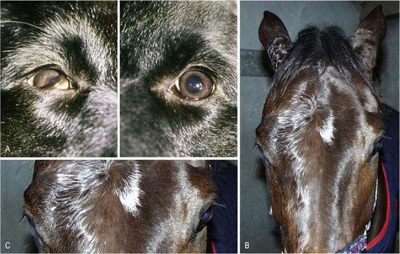
Fig. 12.6 Horner’s syndrome affecting: (A) a dog’s right eye, (B) the right side of the horse’s face. Note the sweating and the drooping of the upper eyelashes (see enlargement, C).
Pupillary function in acute brain disease
Pupil size can be an indicator as to the severity of brainstem damage. Severe bilateral miosis can occur with lesions affecting the forebrain or pretectal area. This may represent increased parasympathetic function due to loss of inhibition of parasympathetic LMNs in the midbrain by UMNs of the forebrain. An expanding lesion in the brainstem can affect CN III function. Experimentally, miosis is caused by compression of the rostral colliculus, whereas mydriasis is caused by compression of the parasympathetic nucleus of CN III, or the proximal portion of CN III as it travels along the floor of the calvarium. Brainstem compression can occur with brain herniation in which neural tissue is forced, rostrally or caudally, under the tentorium cerebelli.
Asymmetrical compression can cause unilateral mydriasis, but severe bilateral compression can cause bilaterally, fixed and dilated pupils.Resolution of the miosis is a favourable prognostic sign but progression from miotic to mydriatic pupils indicates increasing severity of the midbrain lesion.
Pharmacological testing of the pupils
Two principles underlie the use of drugs to determine the site of a lesion in the ANS:
1. Drugs can mimic the neurotransmitters at the synapses. They can stimulate the postsynaptic fibre by mimicking the neurotransmitter in the ganglion where pre- and postsynaptic fibres synapse. Or they can stimulate the effector smooth muscle at the autonomic neuromuscular junction. Cholinergic drugs will stimulate the postsynaptic LMN of both the sympathetic and parasympathetic systems, and the neuroeffector junction in the parasympathetic system. Adrenergic drugs will only act at the sympathetic neuroeffector junction.
2. Denervated tissue is hypersensitive to concentrations of neurotransmitter that normally would not stimulate it. Loss of the presynaptic neuron results in denervation hypersensitivity of the postsynaptic neuron. Loss of the postsynaptic neuron results in denervation hypersensitivity of the smooth muscle.
Using these concepts, the clinician may be able to determine whether the animal’s signs are due to loss of either the pre- or postsynaptic fibre (Table 12.3). A dilute concentration of drug that mimics the neurotransmitter at either the interneural synapse or the neuroeffector junction is applied. It will only reverse the signs if denervation hypersensitivity is present.
Table 12.3 Pharmacological localisation of lesions in ANS innervation of the eye
| Drug and mechanism | Effect and lesion localisation |
| Phenylephrine (10%) or adrenalin (0.001%) - direct acting sympathomimetic | Pupil dilation (dog) or change in eyelash angle (horse) if postsynaptic sympathetic neuron has been lost. Minimal and delayed effect if the presynaptic neuron has been lost |
| Hydroxyamphetamine (1%), indirect-acting sympathomimetic that triggers release of adrenaline at neuroeffector junction | Pupil dilation (dog) if presynaptic sympathetic neuron has been lost |
| No effect if postsynaptic sympathetic neuron has been lost | |
| Pilocarpine (2%), direct acting parasympathomimetic | Rapid pupil constriction if postsynaptic parasympathetic neuron has been lost. Reduced and slower effect if presynaptic neuron has been lost |
| Physostigmine (0.5%), indirect-acting parasympathomimetic that inhibits acetylcholinesterase | Pupil constriction if presynaptic parasympathetic neuron has been lost. No effect if postsynaptic neuron has been lost |