CONFORMATION AND EXTERNAL FEATURES IN CATTLE
The features of the bovine head that first attract notice are the angular, pyramidal form, the bare muzzle, and the horns (when these are present). The form owes much to the late development of the frontal sinuses that invade the bones of the cranial vault, transforming the domed contours of the calf’s head into the broad, flattened forehead and upright nuchal surface of the adult (Figures 25-1, 25-2, and 25-3).
The proportions are also much altered after birth by the greater growth of the facial part than of the neurocranium.The modified skin around the nostrils extends to the margin of the upper lip, forming the slightly cobbled naked nasolabial plate. This is kept moist by the watery secretion of a thick layer of eccrine glands massed below the skin.
The naked integument continues through the large oval nostril into the nasal vestibule where it blends with the mucosa. The opening of the nasolacrimal duct is placed just caudal to the mucocutaneous junction. It is concealed on the ventromedial side of the fold that prolongs the ventral concha rostrally but may be uncovered for cannulation by bending the wing of the nostril outward.
The lips are thick, relatively immobile, and insensitive; they take little part in prehension of food. The upper one is the larger and overlaps the lower lip to the front and sides when at rest.
The size and conformation of the horns depend on breed, age, and sex. The horns are based on much smaller cornual processes that grow from the frontal bones at the caudolateral angles of the forehead. The cornual process has a ridged and porous surface and is covered by a papillated dermis that also serves as periosteum. The specialized dermis blends with that of the surrounding skin at the base of the projection. The major part of the horn wall or sheath grows from the epithelium that covers the dermis over the horn process; the softer outermost layer (epiceras) is produced by an irregular epithelial strip at the base that is transitional to the ordinary epidermis.
The horn sheath represents a modification of the cornified stratum of the epithelium and consists chiefly of tubules formed over the dermal papillae; the tubules run lengthwise and are welded together by irregular, intertubular horn produced by the interpapillary regions of the epithelium. Since the whole epithelial surface is productive and the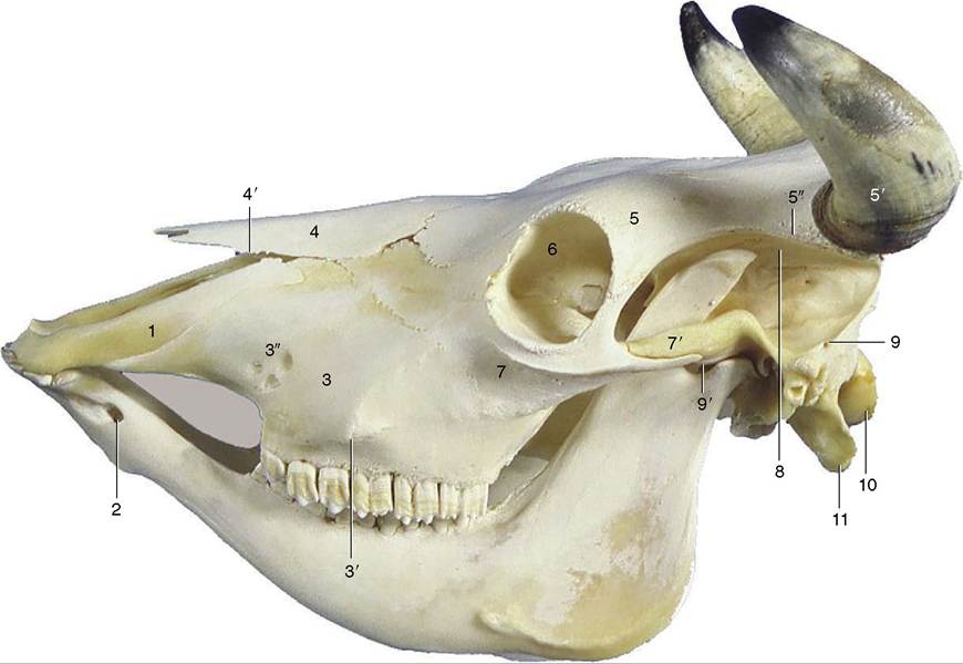
Figure 25-1 Lateral view of bovine skull. 1, Incisive bone; 2, mental foramen; 3, maxilla; 3', facial tuberosity; 3", infraorbital foramen; 4, nasal bone; 4', nasoincisive notch; 5, frontal bone; 5', horn surrounding cornual process of frontal bone; 5", temporal line; 6, orbit; 7, zygomatic bone; 7’, zygomatic arch; 8, temporal fossa; 9, temporal bone; 9', temporomandibular joint; 10, occipital condyle; 11, paracondylar process.
older horn is thrust apically by that of more recent origin, it follows that the horn sheath increases in thickness toward the tip (Figure 25-4). Although horn growth is continuous, the rate of production varies according to the stresses to which the animal is subjected, and it is usual to find the horns marked by alternating rings of greater and lesser thickness. The latter represent periods when production was less active and the horn that was produced was softer and more prone to wear. In cows these periods commonly correspond to calvings. Since the first calf is generally born when the cow is about 2 years of age and subsequent calves are born at yearly intervals thereafter, the number of rings is commonly one fewer than the animal’s age in years (Figure 25-5).
The sensitive dermis of the horn is supplied mainly by the cornual nerve (Figure 25-6/1), a branch of the zygomaticotemporal division of the maxillary nerve. The cornual nerve arises within the orbit and then passes backward through the temporal fossa, where it is sheltered by the prominent ridge of the temporal line.
The nerve later divides into two or more branches that wind around this ridge and approach the horn separately under cover of the thin frontalis muscle. The cornual nerve is often blocked for dehorning operations and is then sought where it crosses the ridge, roughly midway between the postorbital bar and the horn (Figure 25-6/1). The anesthetic technique is not always successful; among the explanations advanced to account for its failure are variation in the relationship of the nerve to the bony ridge, precocious division into divergent branches, and the existence of unusually substantial contributions from the supraorbital or infratrochlear nerves. Since the nerve to the frontal sinus may extend to the diverticulum within the horn, even infiltration around the horn base does not ensure complete loss of sensitivity.The cornual nerve is accompanied by a considerable artery and vein that branch from the superficial temporal vessels within the temporal fossa. The artery ramifies before it reaches the horn. Its smaller branches run in the grooves and canals of the cornual process and retract when severed so that they cannot be easily grasped with hemostats; because of this, dehorning is
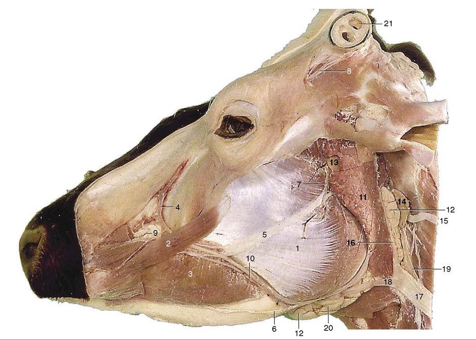
Figure 25-2 Superficial dissection of the head. 1, Masseter; 2, zygomaticus; 3, buccinator; 4, facial vein, 5, 6, dorsal and ventral buccal branches of facial nerve; 7, auriculotemporal nerve; 8, cornual nerve; 9, infraorbital nerve; 10, parotid duct and facial artery and vein; 11, parotid gland; 12, mandibular gland; 13, parotid lymph node; 14, lateral retropharyngeal lymph node; 15, spinal accessory nerve; 16, maxillary vein; 17, external jugular vein; 18, linguofacial vein; 19, common carotid artery; 20, mandibular lymph node; 21, cornual diverticulum of frontal sinus.
accompanied by spurting arterial hemorrhage unless the cut is made close to the skull where the arteries are still embedded in soft tissue.
The horns are barely indicated in the newborn calf, and their development can be prevented by cauterization of the germinal epithelium at an early age (2 to 4 weeks). The epidermis, which spreads to heal the wound, lacks the specialized inductive capacity of the original covering. An extension from the frontal sinus invades the cornual process when the calf is about 6 months old.