CONFORMATION AND EXTERNAL FEATURES IN SHEEP AND GOATS
The shape and appearance of the head show many specific, breed, sex, and age differences, but although they determine the “character” of the animal, they are for the most part of no great clinical interest.
It is, however, important to note that the dorsal profile of the skull, unlike that of adult cattle, is domed over the cranial cavity and slopes caudally toward the nuchal plane; this feature is commonly masked by the location and size of the horns (Figure 25-7).The goat’s head has a fairly long coat of hair, but that of sheep is shorter, and in some breeds wool extends considerably onto the face. The nasal plate resembles that of the dog but has a more limited extent, particularly in goats. It is confined to a narrow strip to each side of the deep median philtrum, with lateral prolongations along the upper edges of the long, slitlike nostrils.
The horns arise close behind the orbits in a parietal position (see Figure 26-2). Each is based on a separate ossification center that makes a secondary fusion to a projection of the skull quite close to its contralateral fellow. In both sheep and goats the frontal sinus later excavates the horn core at the base but does not reach so far toward the tip as in cattle. Polled breeds are common, but when horns occur they are generally present in both sexes, although those of males are more
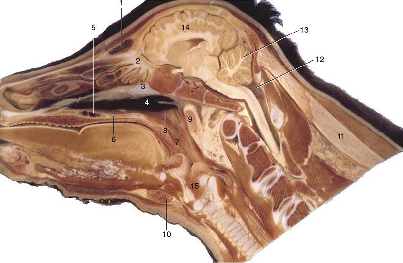
Figure 25-3 Paramedian section of the head of a 2-week-old calf. Note the rounded vault. 1, Frontal sinus; 2, ethmoidal conchae; 3, vomer; 4, pharyngeal septum; 5, palatine sinus; 6, hard palate; 7, soft palate; 8, nasopharynx; 9, medial retropharyngeal lymph node; 10, mandibular gland; 11, nuchal ligament; 12, cerebellomedullary cistern; 13, cerebellum; 14, cerebrum; 15, larynx.
strongly formed.
In a few rare breeds, two (in rams occasionally three) pairs may exist. The multiple-horn (polycerate) condition is frequently associated with defects of cranial sutural closure and also of the eyelids.The horns of goats generally have an oval section and grow caudally over the skull. Those of sheep are triangular in section and pursue a helical course that carries them first caudally, then successively ventrally, rostrally, and dorsally in a form of increasing complexity. This growth sometimes carries the inner surface of the horn close to the skin of the face, which may suffer from pressure necrosis if contact is made. Shepherds of flocks of the vulnerable breeds are on watch for this occurrence and often remove a surface slice from the horn in treatment or in prevention. The operation can be performed without anesthetic if only “horn” is sawn away; on occasion sensitive dermis and bone must also be removed.
The horns of the sheep and goat are placed so close to the orbit that the supplying structures ascend directly behind the zygomatic process, where the nerve may be blocked. The horn of the goat receives a subsidiary supply through branches of the infratrochlear nerve; these can be reached by a second injection at the dor- somedial margin of the orbit.
Certain glands of the skin of the heads of sheep and goats are mentioned in Chapter 10.