Cytoplasm and Cytoplasmic Organelles
Cytoplasm
Cytoplasm is the material filling the inside of the cell and containing the intracellular organelles and nucleus (Fig. 2-18). Organelles are intracellular structures organized for a particular function within the cell.
These include the Golgi apparatus, endoplasmic reticulum (smooth and rough), mitochondria, centrioles, free ribosomes, lysosomes, peroxisomes, and a variety of crystals, granules, and droplets (formerly called inclusions, collectively). The relatively liquid component of cytoplasm is cytosol; the organelles are arranged within the cytosol by a complex system of intracellular filaments and microtubules called the cytoskeleton.Dissolved or suspended within the fluid cytosol are a variety of proteins, sugars, and salts. Many of the proteins are enzymes that function in the metabolic activities of the cell. Within some cells are proteins that function as cytosolic receptors binding ligands that have gained access to the cytosol after passing through the cell membrane.
The Golgi Apparatus
The Golgi apparatus varies in size and location in cells of different tissues but generally appears as a stack of flattened membranous sacs (lamellae) near the nucleus (Fig. 2-18). It functions as the site of the final stages of synthesis and packaging of secretory products of the cell. Within the Golgi, secretory products are enclosed within a membrane vesicle for temporary storage in the cell or for transport to the plasma membrane, where exocytosis releases
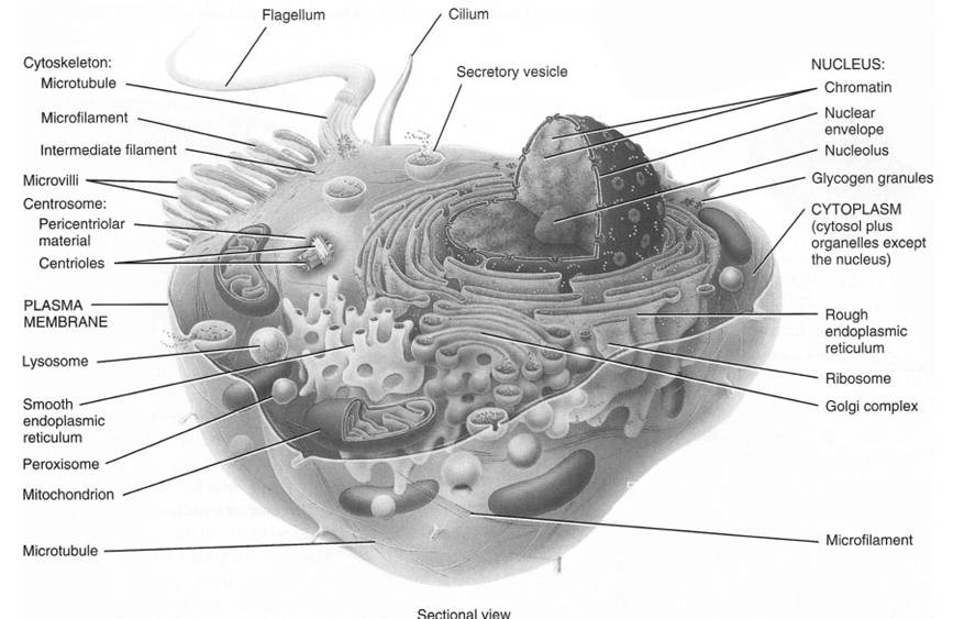
Figure 2-18. General organization of a typical cell. (Reprinted with permission of Wiley-Blackwell from Tortora, G.J. and Derrickson, B., Principles of Anatomy and Physiology, 11th ed. Hoboken, NJ: John Wiley & Sons, 2006.)
the products into the extracellular fluid as a form of secretion.
Mucopolysaccharides may form in the Golgi apparatus, and glycoproteins are terminally synthesized there as combinations of carbohydrates and proteins that have been transported to the Golgi apparatus by the smooth and rough endoplasmic reticulum.The Endoplasmic Reticulum and Ribosomes
The endoplasmic reticulum is a membranous network found throughout the cytoplasm of the cell (Fig. 2-18). It was first described in the endoplasm (the cytoplasm deepest in the cell), giving rise to the name endoplasmic reticulum. Although still called the endoplasmic reticulum, it has been observed in all parts of the cytoplasm and may be continuous with the outer nuclear membrane. The endoplasmic reticulum is in the form of tubules and sheets, with occasional enlarged sacs or vesicles called cisternae.
in some sites, the endoplasmic reticulum is associated with ribosomes, which appear like beads along the membrane. Granular or rough endoplasmic reticulum (Fig.2-18) is endoplasmic reticulum associated with ribosomes; agranular, or smooth endoplasmic reticulum (Fig. 2-18), has no ribosomes associated with it.
Mitochondria
Mitochondria are ovoid organelles about 10 μm long. The double membrane of the mitochondrion, with the cristae projecting into the interior, provides a large surface area for attachment of enzymes (Fig. 2-18). Studies of fragmented mitochondria indicate that all of the enzymes associated with oxidation of nutrients to carbon dioxide, ATP, and water are found in the mitochondria. Thus, all of the enzymes and coenzymes involved in the tricarboxylic acid cycle (also called the Krebs cycle, or citric acid cycle) are largely local to the mitochondria.
oxidation during the tricarboxylic acid cycle releases carbon dioxide and hydrogen atom pairs (H2). The H2 furnishes its electrons to the mitochondrial electron transport system to drive a series of reduction reactions, culminating in the formation of water and storage of the energy in the form of ATP.
The ATP is formed by the oxidative phosphorylation of ADP (adenosine diphosphate), which adds one inorganic phosphate molecule to ADP, creating a higher- energy compound. The energy incorporated into ATP becomes available for any cellular activity that requires energy, such as protein synthesis, muscle contraction, and active transport. Energy is released during the reconversion of ATP to ADP and an inorganic phosphate. Most of the cellular processes requiring energy take place outside of the mitochondria.since the mitochondria produce the energy for the cell, it follows that the more mitochondria in a cell, the more active the cell can be. Mitochondrial ATP production depends on oxygen, so highly active cells also need a ready supply of oxygen. The enzymes of the glycolytic pathway, using glucose as a substrate, can produce ATP without oxygen in the cytoplasm. However, this pathway is less efficient than mitochondrial production; it produces less ATP per molecule of substrate.
Mitochondria contain their own DNA and RNA for reproducing themselves. They also carry on partial synthesis of proteins and lipids and have their own ribosomes. Mitochondrial reproduction can be stimulated by increased demands for cell energy and is not dependent on cellular division.
Lysosomes
Lysosomes are membrane-bound vesicles of digestive (hydrolytic) enzymes. They are larger than ribosomes but smaller than mitochondria, ranging in diameter from 0.25 to 0.75 μm. Lysosomes apparently originate from the endoplasmic reticulum and the Golgi apparatus.
Lysosomes contain a variety of enzymes that degrade all types of biologic molecules. Normally, the membrane of the lysosome prevents lysosomal enzymes from acting on molecules within the cytoplasm. However, in certain conditions, the enzymes are released into the cytosol, which may then lyse (destroy) the cell itself.
Cytoplasmic vesicles formed by the phagocytosis of extracellular material may fuse with lysosomes, thereby permitting the enzymatic digestion of the contents of the vesicle while protecting the cell itself from lysis.
White blood cells, which act as scavenger cells by phagocytizing bacteria, dead tissue, and damaged cell debris, contain many lysosomes. Lysosomes also engulf and degrade intracellular organelles. This is a means by which individual cells can remove and recover components of damaged parts of themselves. The only mammalian cells that are known not to contain lysosomes are the red blood cells.When cells contain inactive lysosomes, disease can follow. An example is Pompe’s disease, in which the lysosomes cannot digest glycogen. Also, changes caused by sunburn occur when the ultraviolet light of the sun ruptures lysosomes in the skin cells.
Other Structures
Peroxisomes are smaller than lysosomes and are most numerous in cells of the liver and kidney. Peroxisomes contain enzymes responsible for degrading lipids, alcohols, and a variety of potentially toxic substances. Hydrogen peroxide is produced within the peroxisome as a result of this enzymatic action. other enzymes within the peroxisome rapidly degrade the potentially toxic hydrogen peroxide to protect the cell.
Microtubules, intermediate filaments, and microfilaments are rodlike organelles that make up the cytoskeleton, which primarily functions to determine the shape of the cell and assist with cell movement. Microtubules are scattered throughout the cytoplasm in most cells and are
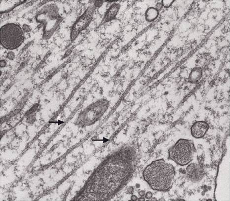
Figure 2-19. Microtubules in axons of neurosecretory neurons. (Reprinted with permission of Wiley-Blackwell from Dellmann, HD and Eurell, J. Textbook of Veterinary Histology. 5th ed. Baltimore: Lippincott Williams & Wilkins, 1998.)
the largest and most rigid of the three cytoskel- etal components (Fig. 2-19). Microtubules are spindle fibers in cell division, motile elements in cilia, and assisters of transport of molecules within some cells, such as in the processes of neurons (nerve cells). intermediate filaments are primarily found in association with specialized cell-to-cell junctions, such as desmosomes (Fig. 2-9).
Microfilaments are thinner than microtubules, but they make up most of the cytoskeleton. Microfilaments are composed of actin, a protein involved in cell movement and muscle contraction.
The centriole is a short cylinder about 0.2 μm wide and 0.4 μm long. Centrioles, composed of nine triplets of microtubules, usually occur at the bases of cilia, where they are called basal bodies. A pair of centrioles, the centrosome, also occurs in all cells near the nucleus and organizes the microtubules, which form the mitotic spindle during cell division.