Dentition
The mammalian dentition* possesses certain characters that in combination, if not individually, are diagnostic of the class. The complement of teeth is limited to a fairly small number, rarely exceeding 44 in the permanent dentition, which is determined for each species — although minor variations may occur.
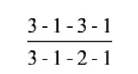
Unlike those of most other vertebrates, the teeth of mammals are very differently developed in different regions of the mouth for better performance of special tasks; this character, known as heterodonty, allows the recognition of incisor, canine, premolar, and molar groups. A single replacement of the teeth first erupted is provided by a second, stronger set that is better adapted to the larger jaws and to the more vigorous mastication of the adult. The sequence is known as diphyodonty, in contrast to the polyphyodonty (multiple succession) in most other vertebrates. Finally, the teeth are implanted in sockets set along the margins of the jaws, an arrangement described as thecodont.The number and classification of the teeth in a particular species are conveniently represented by a formula in which I stands for incisor, C for canine, P for premolar, and M for molar. For the dog, the formula of the permanent dentition may be written


without risk of confusion, as molar teeth are always lacking in the milk set. There are various notations for the identification of individual teeth. The most convenient would use upper- and lowercase letters to denote the permanent and temporary, respectively, and superscript and subscript numerals to show the upper and lower teeth, respectively.
For example, P1 may stand for the first permanent upper premolar, i2 for the secondary temporary lower incisor.The term diastema is used for a considerable gap between teeth in the one jaw, most usually for that between the incisors and premolars.
The description of a simple tooth may be considered before the discussion returns to the features of the different types of teeth. A tooth (dens) consists of crown and root, and each is easily distinguished. The crown is encased in enamel, a very resistant, calcified, slightly opalescent, white
material, whereas the root is encased in cement, a softer, less shiny, yellowish tissue. The part of the tooth between root and crown is termed the neck (Fig. 3.13). Certain variations in structure may occur at the neck: the cement and enamel commonly abut, but the cement may overlie the enamel or sometimes the two tissues fail to meet, exposing a narrow strip of dentin, the third calcified tissue of the tooth. The dentin, which is also known as ivory, provides the greater part of the substance of the tooth and contains a small central cavity that houses the connective tissue pulp. The pulp continues through a canal in the root of the tooth to merge with the connective tissue in the depth of the tooth socket (alveolus).
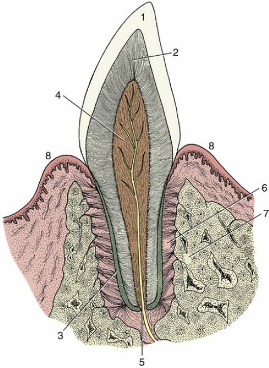
FIG. 3.13 Schematic longitudinal section of a simple tooth. 1, Enamel; 2, dentin; 3, cement; 4, pulp; 5, apical foramen; 6, periodontal ligament; 7, socket (alveolus); 8, gum.
Fig. 3.13 depicts the idealized condition in which both the gum (gingiva) embraces the neck and the crown corresponds to the exposed part of the tooth. The gums may recede with advancing age, exposing the cervical part of the root, a condition familiar in many older people who are said, on this account, to be “long in the tooth." In many mammals, especially the herbivores, part of the enamel-covered crown is concealed below the gum line, which is extruded gradually to compensate for the loss at the masticator surface.
Such high-crowned teeth are called hypsodont (or hypseledont) and are characteristic of animals feeding on abrasive food. Even in species such as primates or dogs with low-crowned (brachydont) teeth suited to a softer diet that produces less wear, part of the enamel-covered region commonly lies below the gum during initial use of the tooth. For these reasons it is useful to distinguish the “clinical crown" from the anatomic crown: the first term specifies the exposed part of the tooth regardless of its structure, and the second specifies the enamel-covered part regardless of its location (Fig. 3.14).The detailed description of the crown requires some system for indicating its various surfaces because the usual terms of relative position are inadequate, considering that the curved line followed by the tooth row (arcade) alters the orientation of equivalent surfaces of successive teeth in the series. Less ambiguous terms are vestibular (labial, buccal) and lingual and mesial and distal; their usage is indicated in Fig. 3.18. Where adjacent teeth touch, the appropriate mesial and distal surfaces may both be termed contact surfaces. The working area, if extensive and not a mere cutting edge, is known as the occlusal or masticatory surface.
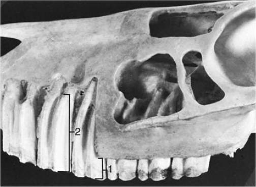
FIG. 3.14 Premolar teeth exposed in the upper jaw of a horse. The part protruding above the gum is the clinical crown (1); the whole enamel-covered part is the anatomic crown or body (2) of the tooth.
Enamel is a densely calcified tissue of ectodermal origin. It is acellular and lacks regenerative capacity to patch a hole or repair a fracture. Because it is exposed to rough treatment, it is necessarily very hard. Nevertheless, the enamel casing may eventually be breached, and the softer dentin that wears away more rapidly would then be exposed. The thickness and the resistance of the enamel therefore largely determine the working life of the brachydont tooth.
In species in which the tooth crown is high and only gradually passed above the gum line, the enamel may be folded in a very complicated fashion; this folding increases the efficiency of the masticatory surface, because the unequal resistance of the tissues exposed on opening of the enamel casing results in an irregular ridged arrangement (see Figs. 3.19 and 18.20).Cement is the least hard of the calcified tissues of the tooth and resembles bone in structure, although it lacks so regular an organization. The initial deposit over the root is thin, but as deposition continues throughout life it may eventually form quite a thick crust. Collagen fibers extend from the cement into the periodontal ligament or membrane (periodontium), the specialized connective tissue that fastens the tooth in its socket. Although broadly comparable to bone in structure and development, cement differs in one important respect: it is relatively immune to pressure erosion. Orthodontists make use of this characteristic when they adjust the position of a tooth in the jaw by fitting an appliance that presses the tooth against the alveolar wall. If the adjustment is performed correctly, the pressure produces an erosion of the bone but leaves the tooth unaffected and free to shift into the space created. This lack of response to pressure is relative, not absolute, and excessive pressure causes resorption; indeed, the roots of the temporary teeth are resorbed under pressure from their permanent replacements thrusting against them.
Dentin is also similar to bone in having a calcified, collagen-rich matrix. In bone the osteoblasts become imprisoned in the matrix, but the dentin-producing cells (odontoblasts) recede from the newly formed dentin and remain as a continuous layer on the surface lining the dental (pulp) cavity. The odontoblasts retain their productive capacity throughout life, and a slow but continuous production of secondary dentin, with corresponding reduction of the dental cavity, continues into old age.
This process may be accelerated when local damage or abrasion of the crown threatens to expose the pulp. Secondary dentin is easily recognized from its darker color. Although once disputed, it is generally believed that fine nerve processes enter a short distance into the dentin from the pulp.The dental cavity reflects the external form of the tooth, sending a branch into each major elevation of the crown and through a narrow passage in the root where it opens at the apical foramen. There may be more than one root, with each joining the central cavity via a channel.
The pulp that fills this space is a very delicate connective tissue margined by the odontoblast layer and richly vascularized. A lymphatic plexus also exists, although it is difficult to demonstrate. Numerous nerves run within the pulp; some are vasomotor, although most are sensory and possess endings that can be stimulated in various ways. Whatever the stimulus, thermal, mechanical, or chemical, the sensation perceived is pain. Because the pulp is contained within unyielding walls, even a slight inflammatory swelling is quickly appreciated.
Each tooth is implanted in a separate socket in the margin of a jaw. The form of the socket corresponds to that of the root and is therefore often branched and irregular. Where the teeth lie close together the septa between adjacent sockets may be very delicate or even defective. Typically, the socket is lined by a thin lamina of compact bone perforated for the passage of the vessels and nerves supplying both the socket and the tooth. The outer surface of the lamina may be braced by trabeculae of spongy bone extending toward the surface of the jaw or radiating into surrounding parts. In the areas where the alveolar margin is narrow, however, the lamina merges with the external compacta of the jaw. The tooth is attached to the socket by means of the tough fibrous periodontal ligament made up of collagen fibers. The fibers are attached to both the cement and the alveolar bone and actually suspend the tooth in a sling.
This arrangement allows the tooth a certain (though usually very limited) mobility, resulting in slight rotation and tilting during mastication.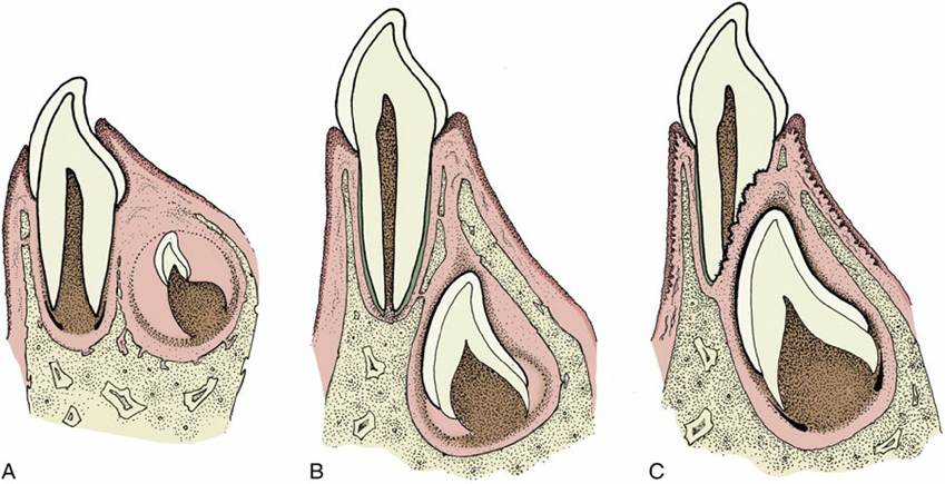
FIG. 3.15 Schematic drawings representing tooth eruption and replacement. (A) Eruption of a deciduous tooth. The primordium of the permanent tooth is located on the lingual side of the deciduous tooth. (B) The fully developed deciduous tooth within a bony alveolus. The crown of the permanent tooth has already formed. (C) The permanent tooth is ready to break through. The root of the deciduous tooth has been resorbed; formation of the root of the permanent tooth is in progress.
The vessels and nerves that supply the teeth are derived from the major trunks (superior and inferior alveolar arteries, veins, and nerves) that course through canals in the jaws.
Tooth eruption is a complicated and controversial process involving a number of factors: root growth, bone growth, pulpal proliferation, tissue pressure, and periodontal traction. Their relative importance is disputed, but the last factor is probably the most significant. The temporary teeth rise in the jaws after the crown is completed but before the root is formed; this process carries the tooth closer to the surface and provides the space necessary for the formation of the root. The movement of the crown is facilitated by a loosening of the connective tissue of the dental follicle and gum and by the presence of remnants of the epithelium of the dental lamina, which define the line of passage. However, if these remnants are large and cystic, as sometimes happens, they may obstruct rather than facilitate the movement of the tooth, divert it from its true path, and give rise to troublesome anomalies of site and spacing. The retention of an epithelial covering over the unerupted crown ensures that no breach of continuity occurs when the tooth breaks through to the surface, as this remnant of the enamel fuses with the epithelium of the gums embracing the tooth
(Fig. 3.15).
The eruption of the permanent teeth is more complicated. They develop in bony crypts deep to the roots of the equivalent teeth of the temporary set. To erupt they must escape from this confinement and displace their predecessors. The erosion of the roof and the continuous adjustment of the walls of the embedded alveolus involve the usual processes of bone remodeling such that the permanent tooth and its alveolus migrate as a unit through the jaw to enter the alveolus of the temporary tooth. The replacement tooth then presses on the root of the temporary tooth, causing its resorption. The attachment of the temporary tooth is loosened, allowing it to shift and become increasingly mobile during mastication, followed by shedding and replacement by the permanent tooth. Proper eruption of the permanent teeth depends on places being held ready for them by the temporary teeth. If the decidual teeth are prematurely lost, the filling of the alveoli by bone may make it difficult for the permanent teeth to establish their proper occlusal relationships.
The dentition of the dog, although relatively simple, is well adapted to the feeding habits of the animal (Fig. 3.16). The incisor teeth are small and peglike and are crowded together in the rostral part of each jaw. On eruption, each upper incisor presents a trilobed crown with a labial cutting edge. The lower incisors are bilobed. These features are lost as wear reduces the tooth to a simple prismatic peg. The name incisor suggests that these teeth are used for dividing food before it is taken into the mouth, but in this species a second and more efficient shear is provided by teeth farther back in the mouth. The incisors in the dog are employed mainly in nibbling and grooming.
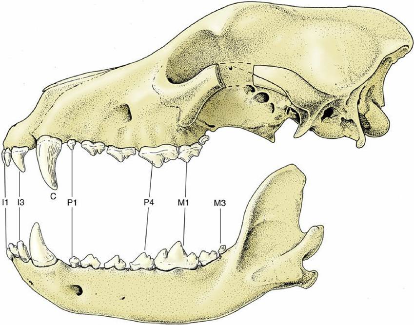
FIG. 3.16 Lateral view of the permanent dentition of the dog. I, incisor; C, canine; P, premolar; M, molar. Numbers indicate position of a tooth, with count beginning at the front.
The canine teeth are particularly well developed, so much so that the generic name (Canis) for doglike animals also is the term for similar teeth in all mammals. Canines are large, curved, and laterally compressed teeth of simple form capable of inflicting a deep wound that are used for aggressive and holding purposes. A large part of each canine tooth is implanted in the jaw. The bony ridge over the alveolus reveals the extent and position of the embedded part of the upper canine.
The premolar and molar teeth together constitute the cheek teeth, a term more common and more useful in describing the assimilation of these two groups of teeth into one in herbivorous species. In all mammals the first few (maximally four) cheek teeth are represented in both dentitions and are assigned to the premolar group; the remainder (maximally three) are represented only in the permanent dentition and are known as molar teeth. The premolars of the dog form an irregular but fairly closely spaced series of increasing size and complexity. The cusps or projections of the individual crowns are aligned one behind the other to form a discontinuous serrated cutting edge to enable a more rapid and cleaner division while the notches help hold the food in place. The more caudal molars also possess a cutting potential but are principally developed for crushing by their broader and more extensive masticatory surfaces. Their cusps or elevations are arranged in a pattern that is faithfully reproduced on the teeth of all members of the species; their homologues can be recognized, although sometimes only with great difficulty, in the teeth of other mammals.
Most of the cheek teeth, unlike the incisors and canines, have more than one root. Multiple roots, especially if divergent, provide firmer anchorage but make extraction difficult.
The dentition of the cat is reduced to
in the permanent set (Fig. 3.17). It is even more closely adapted to a fleshy diet, and crushing potential is largely eliminated with a reduced molar series. The cutting action of the cat's cheek teeth earns them the description secodont; the dual-purpose structure of the dog's molars is better described as tuberculosectorial. The incisors of cats are remarkably small and the canine teeth relatively large.
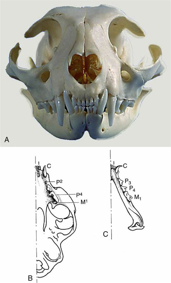
FIG. 3.17 Permanent dentition of the cat. (A) Rostral view. (B) Upper jaw. (C) Lower jaw. I, incisor; C, canine; P, premolar; M, molar. Numbers indicate position of a tooth, with count beginning at the front, with superscript indicating upper jaw and subscript lower jaw.
In other domestic species, the diet is much more abrasive and requires considerably more crushing and grinding. Only the most conspicuous features of the modified dentition are presented here (details in the later chapters). In the dentition of the pig the broad crowns of the cheek teeth carry an elaborate formation of blunt cusps that make them very effective crushing instruments, and these teeth are said to be bunodont (Fig. 3.18). The canine teeth of this species remain open at the embedded end (root) so that accretion of dental tissues continues throughout the animal's life. This persistent growth, coupled with their curved form, allows them to assume very striking forms in older individuals, particularly in boars.
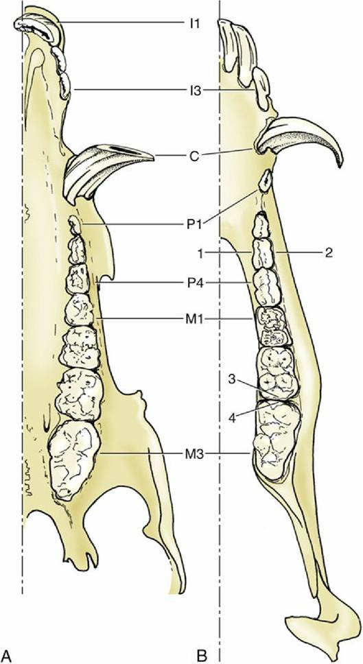
FIG. 3.18 Permanent dentition of the pig. (A) Upper and (B) lower jaws. 1, Lingual surface; 2, vestibular surface; 3, distal surface; 4, mesial surface. I, incisor; C, canine; P, premolar; M, molar. Numbers indicate position of a tooth, with count beginning at the front.
The dentition of horses and ruminants that are restricted to herbivorous diet, in contrast to the diet of the omnivorous pig, must allow for continuous and considerable wear at the masticatory surfaces. This requirement is met by the enlargement of these surfaces, by the increase in height of the crowns, which are only gradually extruded (the delayed development of the roots allows growth to continue for some years after the teeth have come into wear), and, above all, by complicated folding of the enamel. This folding has two important consequences: there is an increased amount of the hardest and most durable component of the tooth that is exposed to reduce its attrition, and alternating harder and softer materials wear at different rates to produce an uneven and rasplike masticatory surface (Figs. 3.19 and 3.20).
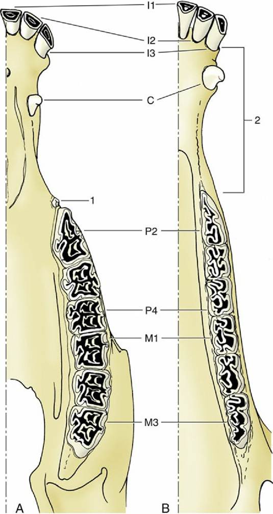
FIG. 3.19 Permanent dentition of the horse. (A) Upper and (B) lower jaws. 1, Wolf tooth (P1); 2, diastema. I, incisor; C, canine; P, premolar; M, molar. Numbers indicate position of a tooth, with count beginning at the front.