DESCENT OF THE TESTES
1. What structures compose the spermatic cord?
2. Read and understand the relationship of scrotal hernias.to the visceral and parietal vaginal tunics.
3. What are cryptorchid testes?
It is helpful to describe the lining of the scrotum and covering of the testis in more detail because it explains the origin of scrotal or inguinal hernias frequently encountered in pigs.
During embryonic development the testes are intraabdominal but outside the peritoneum. They have not yet entered the scrotum, but each has a fibrous connection to the scrotum known as the gubernaculum testis. As development and growth progress, the gubernaculum testis “pulls” the testes through the inguinal canal into the scrotum that creates a double-walled tube of peritoneum. The testis, epididymis, ductus deferens, and testicular vessels, nerves, and lymphatics are enveloped by the inner tube of peritoneum known as the visceral vaginal tunic. The vessels, nerves, lymphatics, and ductus deferens are the components of the spermatic cord (see Figure 14-6). The cremaster muscle (an extension of the internal abdominal oblique muscle) lies on the spermatic cord and assists with drawing.the testes closer to the abdominal wall. The outer tube of peritoneum is known as the parietal vaginal tunic and lines the scrotum (Figure 14-7). The testis and epididymis that are enveloped within the visceral vaginal tunic completely fill the scrotal cavity lined by the parietal vaginal tunic so that only a narrow space remains between the two tunics (the vaginal cavity). The vaginal cavity is continuous with the peritoneal cavity at the vaginal ring (the location where the parietal vaginal tunic of the scrotum is continuous with the parietal peritoneum of the peritoneal cavity). The testicle and vaginal tunics pass through the superficial and deep inguinal rings into the inguinal canal. If the inguinal rings are too large, loops of intestine may enter the vaginal cavity to constitute what is known as an inguinal hernia. An inguinal hernia that has passed into the scrotum is known as a scrotal hernia. The herniated intestinal loops have the potential for strangulation (a cut-off blood supply) or for evisceration (removal from the abdominal cavity) at the time of castration.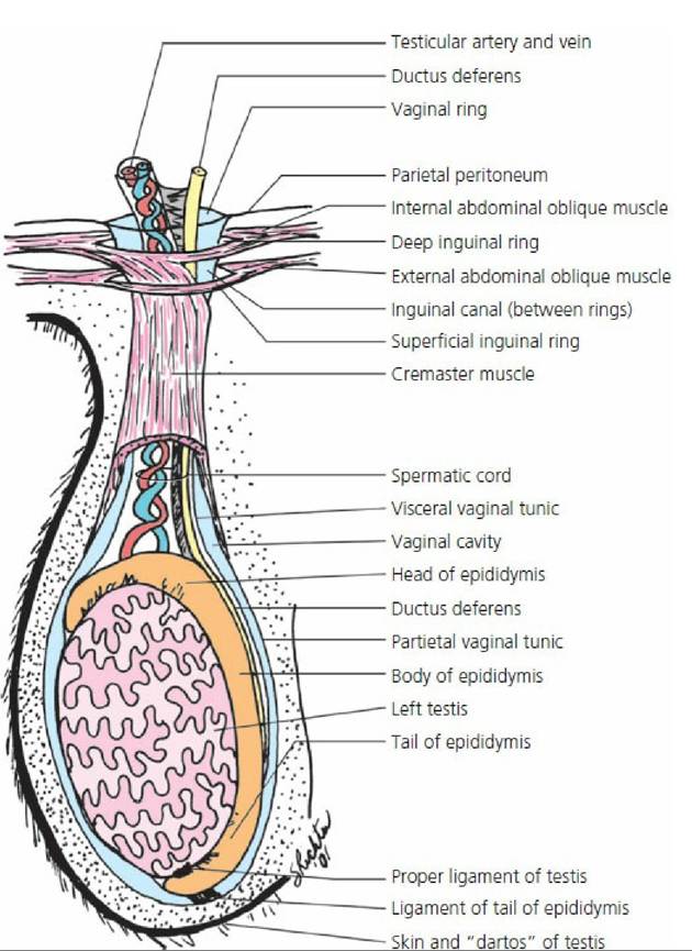
■ FIGURE 14-7 The descended adult testis featuring its relationship to the enveloping visceral vaginal tunic, spermatic cord, the inguinal canal, deep and superficial inguinal rings, vaginal cavity, and peritoneal cavity. The vaginal ring is the location where the parietal vaginal tunic of the scrotum is continuous with the parietal peritoneum. The proper ligament of testis and ligament of tail of epididymis are remnants of gubernaculum testis.
Cryptorchid testes are those that fail to descend. This condition seems to be most prevalent in pigs and horses. When the testis is in the inguinal canal, but not in the scrotum, the horse is referred to as a high flanker. Often the testis or testes are retained entirely within the abdominal cavity.
■