TESTES AND ASSOCIATED STRUCTURES
1. What are the seminiferous tubules?
2. Know the relative location of the Sertoli cells. Are they within the seminiferous tubules?
3. Know the relative location of the Leydig cells.
Are they within the seminiferous tubules?4. Which compartment of the seminiferous tubule provides a home for the spermatogonium? What must it move through to get into the other compartment? What is the name of the compartment where spermatozoa are finally formed?
5. What are the parts of the epididymis?
6. What is accomplished by storage of spermatozoa in the epididymis?
The two testes produce spermatozoa. Although they vary somewhat in size, shape, and location among species, they share a similar structure. The bull testicle and its associated genitalia are shown in Figures 14-1 and 14-2. The seminiferous tubules are convoluted and occupy the greatest portion of each testicle. The spermatozoa are produced within them. The testicle is surrounded by a connective tissue capsule called the tunica albuginea. Support of the seminiferous tubules is provided by connective tissue extensions (septa or trabeculae) into the testis from the tunica albuginea. A cross -section of the testicle (Figure 14-3) shows the relationship of the seminiferous tubules to each other and to their connective tissue support (interstitial tissue).
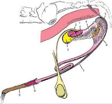
■ FIGURE 14-1 Genital organs of the bull. 1, Seminal vesicle; 2, ampulla of vas deferens; 3, bladder; 4, urethral muscle surrounding pelvic urethra; 5, bulbospongiosus muscle; 6, ischiocavernosus muscle; 7, retractor penis muscle; 8, glans penis; 9, preputial membrane and cavity. (From Roberts SJ. Veterinary Obstetrics and Genital Diseases [Theriogenology]. 3rd edn. Woodstock, VT: Stephen J. Roberts, 1986.)
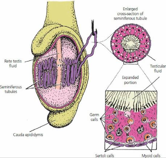
■ FIGURE 14-2 Detailed structure of the testicle.
Only two of the many seminiferous tubule loops are shown. Testicular fluid is secreted by Sertoli cells into the lumen of the seminiferous tubules. Myoid cells are contractile cells contained within the basement membrane. (From Hafez ESE, Hafez B. Reproduction in Farm Animals. 7th ed,. Baltimore, MD: Lippincott Williams & Wilkins, 2000.)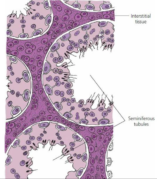
■ FIGURE 14-3 Relationship of the seminiferous tubules to each other and to the interstitial tissue. The interstitial tissue is occupied not only by the usual blood vascular network but also by Leydig cells (interstitial cells) and by connective tissue septa (provides support for seminiferous tubules) from the connective tissue capsule (tunica albuginea) of the testis.
In addition to spermatozoa in various stages of development, two other important cell types are the Sertoli cell (sustentacular cell) and the Leydig cell (interstitial cell). The Sertoli cell provides a “nurse” function for developing spermatozoa. Processes from Sertoli cells surround spermatids and spermatocytes and provide intimate contact with all stages of spermatozoa production; in this respect they are known as sustentacular (supporting) cells. The arrangement of Sertoli cells and the details of seminiferous tubule compartments are shown in Figure 14-4. The Sertoli cells have their base at the periphery of the seminiferous tubules and extend toward the center. The basal junction (tight junction) with adjacent Sertoli cells forms a blood-testis barrier that permits control of the environment within the tubule and also prevents spermatozoa from entering the interstitium. The Sertoli cells divide the seminiferous tubules into two compartments: (1) the basal compartment, which communicates with interstitial fluid and provides space for germinal epithelial cells, and (2) the adluminal compartment, which is the space between Sertoli cells that communicates centrally with the lumen of the tubule.
Division of a germinal epithelial cell (spermatogonium) in the basal compartment provides a replacement cell and another cell, which must move through the Sertoli cell junction to enter the adluminal compartment. Here, further divisions occur and spermatozoa are finally formed. The Sertoli cells secrete a fluid into the adluminal compartment; its composition favors the developing spermatozoa. The Leydig cells are found in the connective tissue surrounding the seminiferous tubules and are responsible for testosterone production.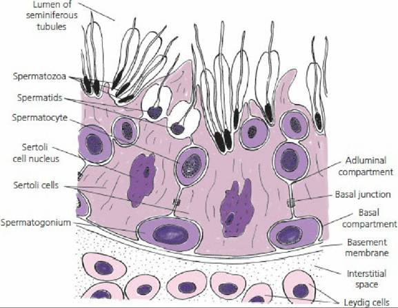
■ FIGURE 14-4 Schematic representation of the periphery of a seminiferous tubule. The Sertoli cells divide the seminiferous tubule into adluminal and basal compartments at their basal junction (tight junction). Leydig cells are in the interstitial space. The basal junction forms a blood-testis barrier whereby the tubule environment is controlled and spermatozoa are prevented from entering the interstitium.
Epididymis
The epididymis is a collection and storage tubule for the testis (Figure 14-5). It begins at the pole of the testis in which blood vessels and nerves enter; this is known as the head of the epididymis. The head continues along one side of the testis as the body of the epididymis, which terminates before making a turn upward as the tail of the epididymis. The head of the epididymis receives sperm and adluminal fluid through efferent ducts from the rete testis (the intratesticular network of straight tubules that receives content from the convoluted seminiferous tubules). Spermatozoa move to the epididymis by the flow of fluid into the lumen of the seminiferous tubules from the adluminal spaces. Storage in the epididymis allows the spermatozoa to reach maturity and become motile. Reabsorption of much of the seminiferous tubular fluid occurs in the head of the epididymis.
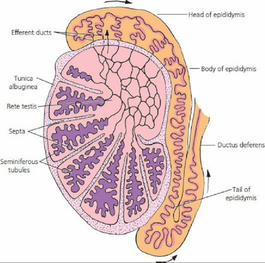
■ FIGURE 14-5 Relationship of the seminiferous tubules to the rete testis, efferent ducts, epididymis, and ductus deferens.
The rete testis is a network of straight tubules connecting convoluted seminiferous tubules with the highly convoluted epididymal tubule via efferent ducts (extratesticular). The flow of spermatozoa with their fluids is shown by the arrows.Ductus Deferens
The ductus deferens (see Figure 14-1), sometimes called the vas deferens, is the continuation of the duct system from the tail of the epididymis to the pelvic urethra. As the ductus deferens leaves the testis, toward the abdomen, it is enclosed along with the testicular artery, vein, nerve, and lymphatic vessels within the visceral layer of the vaginal tunic. This combination of structures is known as the spermatic cord (Figure 14-6). The visceral layer of the vaginal tunic also envelops the testis and epididymis. It is derived from abdominal peritoneum of embryonic origin when the testes descended to the scrotum via the inguinal canal. The inguinal canal is an oblique passage from the abdominal cavity to the exterior of the body that extends from the deep (interior) inguinal ring to the superficial (exterior) inguinal ring. The inguinal rings are slits in the tendinous attachments of the two flat abdominal muscles to the pelvis. After the spermatic cord passes through the inguinal rings, the ductus deferens separates from the spermatic cord to proceed to the pelvic urethra (see Figure 14-1). The ductus deferens terminates with an enlarged, glandular area (variable size among species), known as the ampulla of the ductus deferens (absent in the boar and dog). The relationship of the terminal ductus deferens to the urinary bladder, accessory glands, and pelvic urethra is also shown in Figure 14-1.
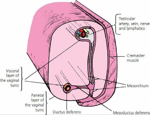
■ FIGURE 14-6 Cross-section of the spermatic cord of mammals. (From Frandson RD, Wilke WL, Fails AD. Anatomy and Physiology of Farm Animals. 7th edn. Ames, IA: Wiley-Blackwell, 2009.)
Scrotum
The scrotum is a cutaneous sac containing the testes. The scrotum contains a subcutaneous layer of smooth muscle fibers, the tunica dartos, which contracts in cold weather and holds the testes closer to the abdominal wall. The scrotum is lined with the parietal layer of the vaginal tunic, which is a continuation of parietal peritoneum into the scrotum.
■