DEVELOPMENT OF THE FEMALE REPRODUCTIVE ORGANS
The initial stages of gonadal development resemble those described for the male. Later, the cell cords fragment into cell clusters, each enclosing an immigrant germ cell. The cords penetrate less deeply into the interior of the gonad than in the male.
The primordial follicles are formed here. Rete formation is less pronounced in the ovary, and because no connection is established with mesonephric tubules, no uninterrupted tubular outlet for the escape of gametes is created (Figure 5-12).Consequently, follicular rupture releases the female gametes at the surface of the ovary by tissue breakdown, a process made easier by the absence of a thick tunica albuginea. The same feature allows for the formation of further sex cords and the establishment of additional follicles during a large part of prenatal life; indeed in certain species this process may continue for a time after birth. Even so, it ceases eventually, and the number of female gametes is then at its maximum; it is afterward depleted by loss through atresia and, to a much smaller extent, through ovulation. Ovarian descent is very limited in most species, being greatest in the ruminants in which the ovaries shift caudally to the abdominopelvic boundary. The duct system of the female is largely provided by the paramesonephric ducts (Figure 5-12/7), which have only vestigial importance in the male. These ducts first develop by invagination of the celomic epithelium lateral to the mesonephric ducts and secondly by active growth in the direction of the urogenital sinus within the genital folds. In contrast, the mesonephric ducts regress in craniocaudal sequence (Figure 5-13), and only remnants survive within the broad ligaments and in the vaginal wall (ducts of
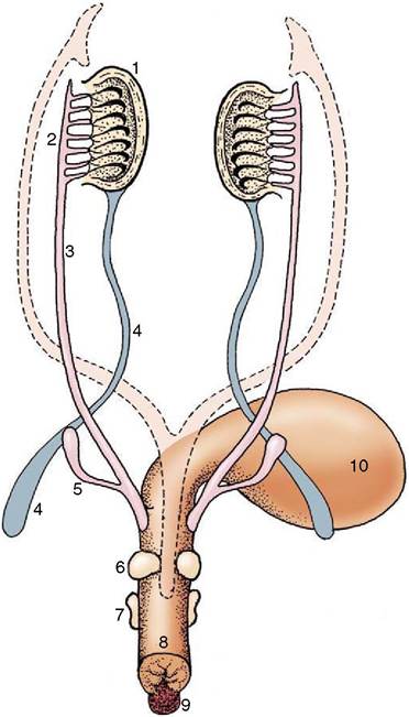
Figure 5-9 Differentiation of the urogenital sinus.
Note the budding of the prostate and bulbourethral glands and the enlargement of the genital tubercle. The regressed parameso- nephric ducts are indicated by the broken lines. 1, Testis; 2, epididymis; 3, deferent duct; 4, gubernaculum; 5, vesicular gland; 6, prostate; 7, bulbourethral gland; 8, urogenital sinus (urethra); 9, genital tubercle; 10, bladder.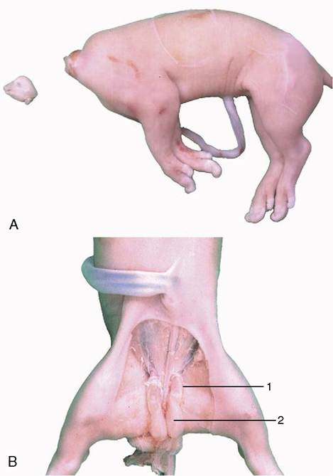
Figure 5-11 A, Pig (fetus) (near term), decapitated in utero 42 days after conception. B, Fetus shown in A with inguinal area dissected to show gubernacula unaffected by removal of pituitary gland. 1, testis; 2, gubernaculum.
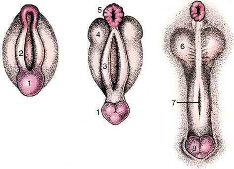
Figure 5-10 Development of the male external genitalia. 1, Genital tubercle; 2, cloacal fold; 3, urogenital fold; 4, lateral (scrotal) swelling; 5, anus; 6, scrotum; 7, groove closing to form the penile urethra; 8, glans penis.
Gartner, ductus epoophori longitudinales), where they are occasionally the seat of anomalous processes. The cranial part of each paramesonephric duct runs lateral to the mesonephric duct, but it crosses this more cau- dally where it inclines to meet and fuse with its fellow (Figure 5-14/d). The cranial end of each parameso- nephric duct remains open to the peritoneal cavity (abdominal ostium of the uterine tube), but the caudal end of the united duct initially ends blindly against a solid outgrowth from the dorsal wall of the urogenital sinus (Figure 5-15). The uterine tubes and the horns, body, and cervix of the uterus form from the parame- sonephric ducts; their caudal parts fuse to an extent that varies with the species and accounts for the very different form and proportions of the uterus of adult animals (p. 199) (Figure 5-16). The supporting genital fold becomes the broad ligament with its various parts. The vaginal lumen appears within the solid outgrowth from the sinus, although a tissue partition, the hymen, may
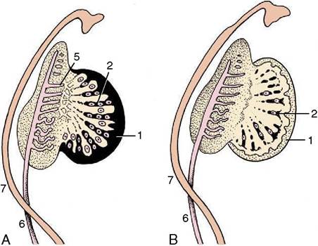
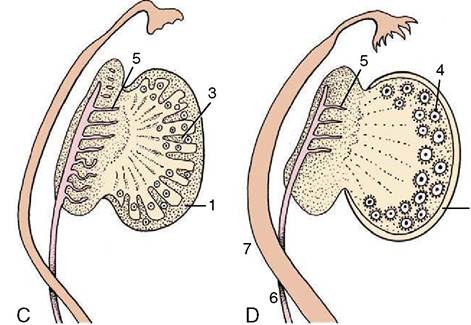
Figure 5-12 Successive stages in the development of the ovary.
1, Celomic epithelium; 2, epithelial cords, penetrating (A) and regressing (B); 3, second formation of sex cords (C); 4, primitive follicles; 5, remnants of mesonephric tubules; 6, mesonephric duct; 7, paramesonephric duct (D).persist near the junction with the fused paramesonephric ducts. A hymen is present only in virgin animals and is rarely well formed in domestic species. Some dispute exists over the contribution of the urogenital and paramesonephric epithelia to the lining of the vagina in the adult, and some suggest that the boundary may divide regions with different responses to hormonal influences that are observed in some species.
The urogenital sinus becomes the vestibule with relatively little further change. Epithelial outgrowths form the vestibular glands in species-variable fashion. The external genital parts are formed from the same structures as in the male; the genital tubercle and lateral folds (swellings) appear first (Figure 5-17). The former produces the clitoris, while the lateral folds, which form the
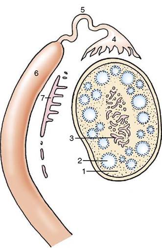
Figure 5-13 Differentiation of paramesonephric duct and regression of mesonephric duct. 1, Interstitial tissue of the ovary; 2, primitive follicles; 3, ovarian rete; 4, infundibulum; 5, uterine tube; 6, uterine horn (4, 5, and 6differentiate from paramesonephric duct); 7, remnants of the mesonephric tubules and duct (epoophoron and paroophoron).
labia majora of human anatomy, regress—with a possible reservation for the bitch. The labia of the vulva of the domestic species are provided by the urogenital folds (Figure 5-17/3) that appear medial to the lateral swellings and correspond to the labia minora of women.