DIFFUSION OF RESPIRATORY GASES
1. Which one of the respiratory gases, O2 or CO2, diffuses more readily through cell membranes?
2. Read the text to understand Table 10-2 and Figure 10-15.
| TABLE 10-2 TOTAL AND PARTIAL PRESSURES (IN MM HG) OF RESPIRATORY GASES IN HUMANS AT REST (SEA LEVEL) | ||||
| GASES | VENOUS BLOOD | Alvelolar air | ARTERIAL BLOOD | TISSUES |
| Oxygen | 40 | 109 | 100 | 30 or less |
| Carbon dioxide | 45 | 40 | 40 | 50 or more |
| Nitrogen | 569 | 564 | 569 | 569 |
| Water vapor | 47 | 47 | 47 | 47 |
| Total | 701 | 760 | 756 | 696 |
| From Reece WO. Respiration in mammals. In: Reece WO, ed. Dukes' Physiology of Domestic Animals. 13th edn. Ames, IA: Wiley- Blackwell, 2015. | ||||
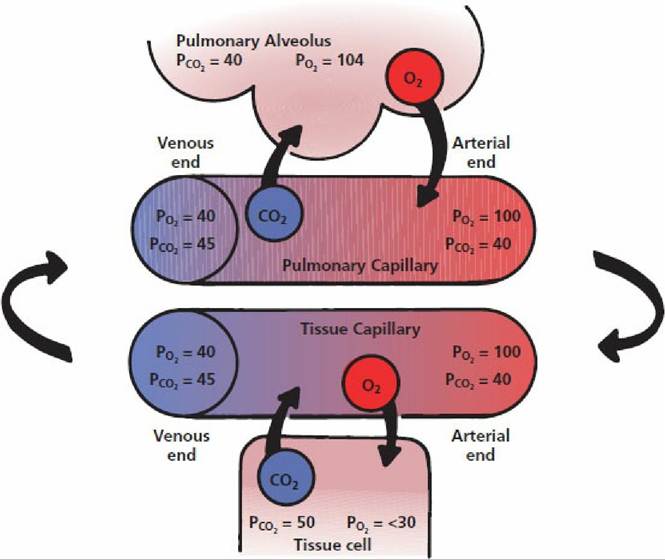
■ FIGURE 10-15 Direction of diffusion for oxygen (O2) and carbon dioxide (CO2), as shown by arrows in progression from the alveoli to the arterial end pulmonary capillaries, arterial end tissue capillaries, tissue cells, venous end tissue capillaries, venous end pulmonary capillaries, and back to the alveoli.
Gas flow as follows: in the pulmonary alveolus the PCO2 is 40 mm Hg and the PO2 is 104 mm Hg; at the arterial end of the pulmonary capillary the PO2 is 100 mm Hg and the PCO2 is 40 mm Hg; at the arterial end of the tissue capillary the PO2 is 100 mm Hg and the PCO2 is 40 mm Hg; in the tissue cell the PCO2 is 50 mm Hg and the PO2 is alveolus to successive solution in interstitial fluid (1), plasma (2), and erythrocyte fluid (3), and finally to combination with hemoglobin (4). The procession of oxygen yield to the cells proceeds in the reverse direction. Diffusion of oxygen to the cells (1) from interstitial fluid and plasma (2) lowers the PO2 of the erythrocyte fluid (3), and just as an increased PO2 increased the saturation of hemoglobin with oxygen, decreased PO2 causes desaturation of hemoglobin (4).Oxygen-Hemoglobin Dissociation Curve
Oxygen dissolves in blood only slightly. If blood contained O2 only in solution, there would need to be about 60 times more blood to transport the 20 volumes % present. Transport is accomplished with the available volume of blood because of the O2 transport potential of the hemoglobin contained in erythrocytes. Oxygen in solution only needs to diffuse into and out of the erythrocytes to be associated with or dissociated from hemoglobin, respectively.
The relationship between the PO2 of blood and the percentage saturation of hemoglobin with oxygen is best described by the oxygen-hemoglobin dissociation curve (Figure 10-17). Note that hemoglobin is nearly 100% saturated when the PO2 of the blood is 100 mm Hg. This is the normal PO2 of arterial blood. Also, at the PO2 of mixed venous blood (about 40 mm Hg), hemoglobin is still about 75% saturated with oxygen. The near 25% that has been lost (dissociated from hemoglobin) corresponds to the utilization coefficient (25/100 = 1/4).
Regardless of the hemoglobin concentration (15 g/dL, normal, or 7.5 g/dL, reduced, as shown in left scales of Figure 10-17), the percentage saturation of hemoglobin is identical for the same PO2 exposure, i.e., the uptake of O2 by hemoglobin (regardless of its concentration) is in equilibrium with the partial pressure of O2. Figure 10-17 illustrates the effect of a reduced hemoglobin concentration on the volume of O2 transported and illustrates that PO2 analysis does not reveal the amount of oxygen present in blood. There would be twice the amount of oxygen in blood having 15 g/ dL hemoglobin than there would be for blood having 7.5 g/dL at any particular PO2. Anemic animals, with low hemoglobin concentrations, may have normal PaO2, but the amount of oxygen in each increment of blood is reduced. To compensate, the heart beats faster to increase blood flow (i.e., more blood (with its reduced oxygen) presented per time period). Figure 10-17 also shows that the rate of oxygen dissociation from hemoglobin increases sharply as the PO2 decrease approaches the middle and lower ends of the PO2 scale. This characteristic of hemoglobin facilitates the provision of oxygen at the capillary level by supplying greater amounts with less lowering of PO2, thus maintaining an adequate pressure difference for diffusion to the cells.