Digestive System
The typical digestive system of a vertebrate such as the cat, consists of a tube with two openings, an anterior mouth and a posterior anus. This tube has become differentiated into a number of specialized areas that we recognize as the organs of the alimentary canal.
The entire alimentary canal is lined by a mucous membrane often exhibiting specialization in various organs. Although the obvious function of a mucous membrane is secretion of mucus, other functions include enzyme secretion, HCl secretion, hormone secretion, etc. Embryological derivatives of the tube,e.g., the liver andpancreas, are physically Integratedintothesystem, usually by meansofducts.There are other organs whose functions are critical to the proper function of the rest of the digestive system. The teeth that figure so prominently in the skull of the skeletal system, the salivary glands that are modified mucous glands, and the tongue all contribute to the proper function of the digestive system.
The main function of the digestive system is mechanical and chemical processing of food into absorbable molecules so that an animal can survive and prosper nutritionally in order to carry out its life processes. Teeth obviously are involved in capturing and holding food and reducing it to chunks that are mixed with salivary secretions containing digestive enzymes and mucus that make it possible for the food to slide down the digestive tract without damaging the lining. The tongue moves the food from the oral cavity into the pharynx and initiates the swallowing reflex. By muscular activity the food is propelled through the regions of the alimentary canal, the pharynx, the esophagus, the stomach, and the small and large intestine. Into the small intestine the secretions of the accessory organs, the liver and pancreas, are added to the secretions of the digestive tract proper.
As the food passes through the alimentary canal it is reduced to a form easily absorbed and made available to all parts of the body. The undigested end product of these processes is fecal material eliminated through the anus.SALIVARY GLANDS AND DUCTS
In order to examine the salivary glands, carefully remove surface connective tissue along the lateral aspect of the head and neck and the surface of the masseter muscle, being especially cautious not to destroy blood vessels, nerves, and nerve-like structures that may be salivary ducts. The parotid gland and its duct are particularly vulnerable to damage while attempting to clean the area below the ear and over the masseter muscle.
The salivary glands are paired and located along the lateral surface of the head beneath connective tissue and skin [Figure 4-1A and Figure 4- 1B]. The largest of these paired glands, located ventral to the ear is the parotid. It is a large, diffuse, lobulated structure that is intimately associated with overlying connective tissue. The parotid duct emerges from approximately the midpoint of the anterior surface of the gland, crosses the masseter muscle, and enters the vestibule (space between the teeth and the inside of the lip) of the oral cavity where it opens opposite the third upper premolar tooth. Two similar appearing bands, one above the parotid duct (the anterior branch of
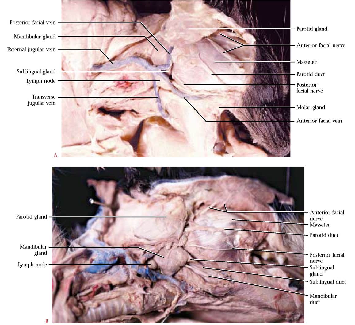
FIGURE 4-1 Salivary glands.
the facial nerve), and one below the parotid duct (the posterior branch of the facial nerve), serve as reference points in identification of the duct [Figure 4-1A and Figure 4-1B].
The mandibular gland also referred to as the submaxillary, is located just ventral to the parotid and posterior to the angular process of the mandible. It, too, is lobular, but the lobes are less diffuse giving the impression that this gland is smoother and more well-defined. Each duct can be identified emerging from beneath the anterior edge of the gland and continuing laterally and beneath the digastric and under the mylohyoid muscles, entering the floor of the oral cavity and opening at the base of a small papilla just anterior to the lingual frenulum [Figure 4-2].
In the vicinity of this gland are generally one or two lymph nodes that can be easily mistaken for the mandibular. These may vary in size and when large may be particularly confusing.The sublingual gland is the smallest of the three salivary glands. It is conical and certainly the smoothest of the three,
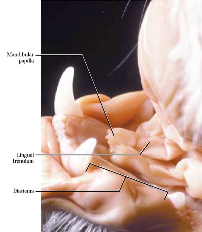
FIGURE 4-2 Mandibular gland papillae.
position of the parotid is similar to that of the cat and its duct (Stensen's) opens in the vestibule opposite the second maxillary molar. The submandibular gland is located medial to the mandible beneath the mucous membrane of the oral cavity and its duct (Wharton's) opens in about the same position as in the cat. The sublingual gland lies anterior to the submandibular along the base of the tongue. Its ducts (Rivinus) open into the floor of the oral cavity along the base of the tongue and directly above the gland. In contrast to the cat, humans do not have molar or zygomatic glands.
ALIMENTARY CANAL
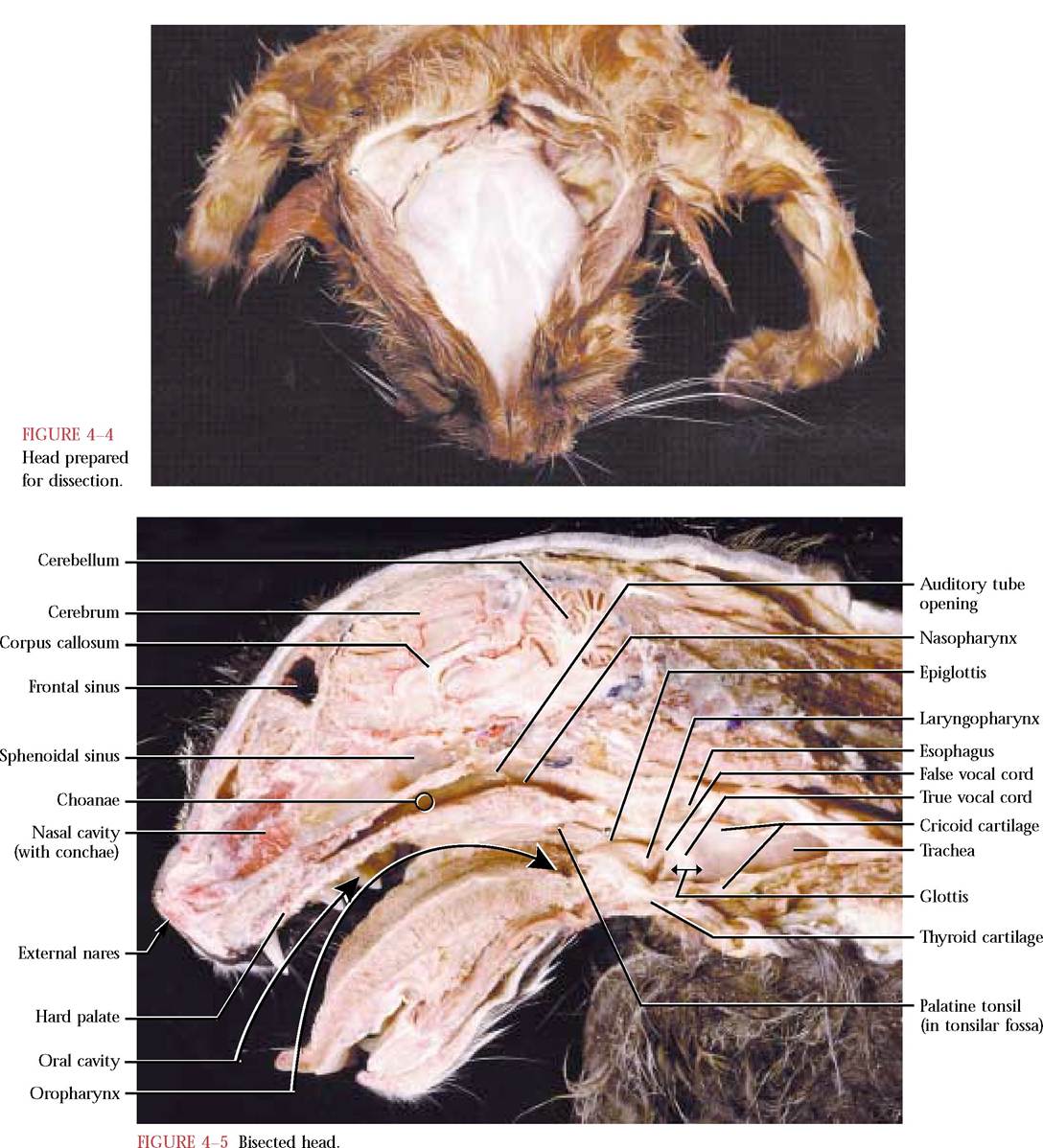
In order to facilitate observation of the anatomy of the oral cavity, the pharynx and the larynx, we will describe two methods of exposing these areas. One of them results in a specimen where the oral cavity, pharynx, and larynx are presented as dorsal and ventral halves while the other is a bisected left and right half. Each possesses its own advantages. The first that we describe is performed with an electrical craniotomy saw and may best be done as a class demonstration. To prepare the cat for this operation, make an incision with a scalpel through the skin from the nose to the back of the neck. Peel the skin back from the incision about 2-2½ inches [Figure 4-4]. The next
and often adheres to the anterior surface of the mandibular gland. This gland wraps around the proximal end of the mandibular duct. The sublingual duct is inconspicuous and runs parallel to the mandibular duct and also opens in the floor of the oral cavity in the vicinity of the mandibular duct [Figure 4-1].
In the cat, two other glands, a molar and a zygomatic, are considered to be part of the salivary system. The molar gland occurs at the angle of the jaw and is located immediately beneath the skin and embedded in the surrounding connective tissue. It has a brownish-gray, granular appearance, and may vary in prominence. Several inconspicuous, small ducts open on the inner surface of the cheek [Figure 4-1A]. The zygomatic or infraorbital gland lies in the floor of the orbit of the eye. It opens by means of a small duct into the posterolateral portion of the roof of the mouth. This gland is difficult to find without removing an eye and we do not recommend that it be done.
In humans, the salivary glands are the parotid, the submandibular (mandibular in the cat), and sublingual. The
steps are probably best performed by the instructor and a laboratory assistant. While the assistant holds the head steady on the dissecting tray, generally locking the thumbs around the ears, and holding either side of the head tightly, the instructor uses the craniotomy saw to bisect the entire head, including the mandible. Both the person doing the cutting and the holding assistant must keep their eyes on the saw at all times! Be sure to wear safety glasses. This is really not as dangerous as it might sound but both individuals should be cautious. The completed bisection may require some minor cutting of the tongue and the laryngeal area with a scalpel. This technique permits viewing of various head organs, e.g., the brain, pituitary gland, nasal conchae, sinuses as well as the relationship of such cavities as the oral and pharyngeal and other difficult to demonstrate organs and openings like the palatine tonsils and the opening of the auditory tube into the nasopharynx [Figure 4-5].
The second dissection is more difficult to perform and should be completed after the salivary glands, ducts, and facial nerves associated with the masseter muscles have been identified. With a sharp scalpel cut through the masseter
muscle to the ramus of the jaw on either side, avoiding the parotid duct and posterior facial nerve, if at all possible.
Use a pair of bone shears to cut through the ramus of the jaw. An audible crunch will be heard and, when completed, should result in your being able to depress the lower jaw. It will be necessary to cut through the juncture of the palatoglossal arches and soft palate to gain full access to the pharyngeal area [Figure 4-6]. This dissection has the advantage of allowing observation of the entire hard and soft palates, the tongue, lingual and labial frenula, opening of the nasopalatine duct, etc. It further avoids some damage to the circulatory and nervous systems that may result from the bisection.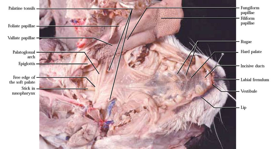
FIGURE 4-6 Dorso-ventrally dissected head.
Mouth or Oral Cavity
The mouth or oral cavity is defined externally by the lips along its border. The lips are a pair of folds whose inner surface is covered by a mucous membrane and whose outer surface is hairy. The space between the lips and the teeth is called the vestibule. In the vestibule, the labial frenulum, a fold of tissue, connects the upper and lower lips at the midline to their respective gumlines [Figure 4-6].
The oral cavity proper is the area of the mouth extending from the lingual side of the teeth to the entrance of the oropharynx [Figure 4-5]. The dental formula of mammals often indicates specializations for dietary habits. In the cat, this formula is:
3incisors: 1 canine: 3premolars: 1 molar 3incisors: 1 canine: 2premolars: 1 molar
The numbers in the numerator of the formula represent the teeth rooted in half of the maxilla and the numbers in the denominator of the formula represent the teeth rooted in one half of the mandible. To determine the total number of teeth, multiply the teeth in each half jaw by two for a total of 30. The teeth of the cat are highly adapted for a carnivorous diet. The small, wedge-shaped incisors are adapted for nipping, the elongated conical canines for stabbing and holding prey and the blade-like molariform teeth for cutting and shearing.
The respective dental formula for humans is:

Humans are described as omnivores, meaning that a combination of plant and animal food is consumed. Therefore, human canines are shorter and blunter and the molariform teeth are flattened and adapted for grinding.
The space between the lower canine and premolars in the cat is called a diastema [Figure 4-2]. Humans do not possess a diastema.
Tongue
The tongue is a very mobile, muscular organ that plays an important versatile role in the life of the cat. It is used as an organ of food manipulation, swallowing, drinking, and grooming. The lingual frenulum anchors the tongue in the anterior floor of the oral cavity and is one of the most obvious structures there. The frenulum becomes obvious when the tongue is lifted [Figure 4-7A].
On the surface of the tongue are four types of projections known as papillae [Figure 4-7B]. The most numerous
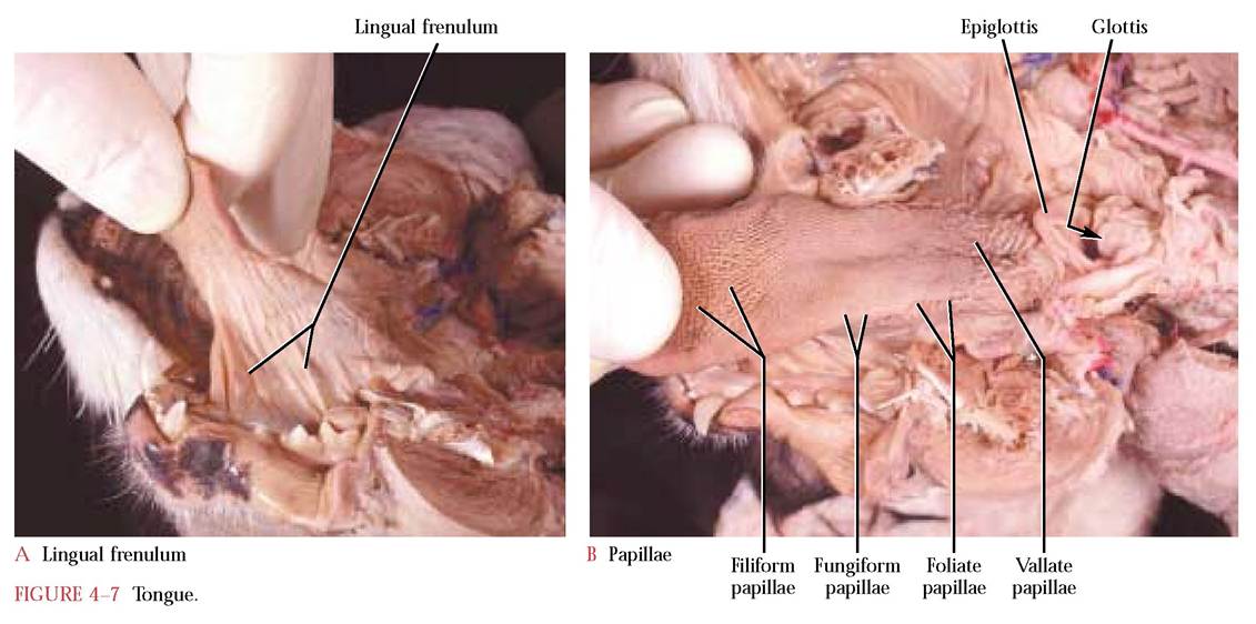
are the filiform, which in cats are located on most of the surface of the tongue. Anteriorly, they appear spiky and are used in grooming or as rasping devices to remove tissue from bones. Posteriorly, they are less pointed. Fungiform papillae are mushroom shaped, less numerous and scattered among the filiform papillae. Vallate papillae are larger, round papillae isolated by shallow grooves and arranged in a V configuration near the root of the tongue. The apex of the V is oriented toward the pharynx. Frequently, these papillae are difficult to distinguish. The foliate papillae are leaf-shaped and are located on the posterolateral aspect of the tongue. Taste buds, microscopic structures important in detecting chemicals identifiable in tasting food, are located in fungiform, vallate, and foliate papillae.
The human tongue is very similar to the cat. Filiform papillae are less acute, vallate are more numerous, and foliate papillae are missing. Fungiform and vallate papillae house the tastebuds.
Palate
The roof of the oral cavity is formed anteriorly by the hard palate and posteriorly by the soft palate. The hard palate consists of a bony shelf constructed of the palatine processes of both the premaxilla and maxilla and the palatines. The hard palate is covered by tissue formed into a series of folds known as rugae. The soft palate consists of connective tissue and muscle and extends from the caudal end of the hard palate to its free edge. At the anterior end of the hard palate and directly posterior to the incisors are a pair of ducts, the incisive ducts, whose openings are distinguished by a small nipplelike structure [Figure 4-6]. These ducts lead to vomeronasal organs whose function amplifies olfaction in mammals who are good smellers. The human palate is similar to the cat with the exception of incisive ducts and reduced rugae. In humans, the absence of the incisive ducts is probably correlated with the reduced sense of smell.
Pharynx
The pharynx, a space shared by the digestive and respiratory systems, extends from the oral cavity to the larynx. It is arbitrarily subdivided into three regions, the nasopharynx, the oropharynx, and the laryngopharynx [Figure 4-5].
The dorsal nasopharynx extends from the internal nares (choanae) to the free edge of the soft palate. Through this passage movement of air for respiration and olfaction occurs. Make a small slit in the soft palate and insert a probe into the nasopharynx or observe this space in a bisected specimen. In the lateral walls of this portion of the pharynx are the paired auditory tube openings, better seen in the bisected specimen [Figure 4-5]. These tubes connect the air filled middle ear cavity with the nasopharynx and are important in equalization of air pressure.
The oropharynx is the space bounded laterally by the palatoglossal arches and extends from approximately the base of the tongue to the free edge of the soft palate [Figure 4-5 and Figure 4-6]. The fauces, the space between the arches marks the transition between the oral cavity and pharynx. Air, food, and liquids pass through the oropharynx on their way to the trachea and the esophagus, respectively. Two small lymphoid masses, the palatine tonsils, lying in
shallow depressions, the tonsilar fossae, are located in the Iaterodorsal walls of the oropharynx [Figure 4-5 and Figure 4-6].
The laryngopharynx is that part of the pharynx continuing from the tip of the epiglottis to the glottis, an opening into the larynx [Figure 4-5].
Esophagus
Movement of food and liquids from the pharynx into the esophagus involves a process called swallowing. During this activity, the larynx is elevated and pulled up against the epiglottis, covering the glottis or opening into the respiratory tract, thereby preventing the food or liquids from entering it and facilitating passage of these materials into the esophagus.
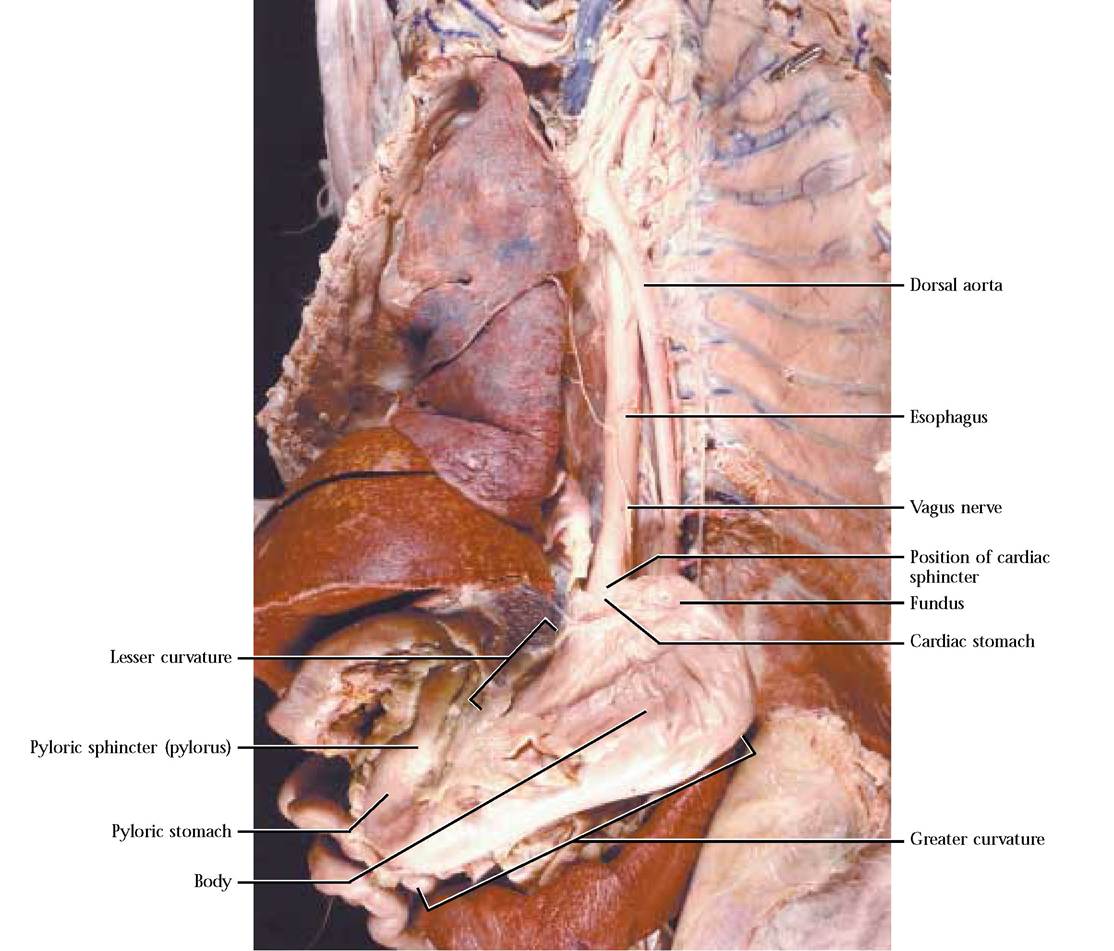
The esophagus is a collapsible muscular tube, capable of being greatly expanded, that passes through the mediastinum dorsal to the trachea. Characteristic wavelike muscular movements (peristalsis) in the walls of the esophagus propel the foodstuff through this tube to the stomach. In mammals, the thoracic and abdominopelvic cavities are separated by a muscular partition known as the diaphragm that plays an essential role in breathing and through which the esophagus passes. At its distal end, the esophagus terminates in a ring of smooth muscle, the cardiac sphincter, permitting movement of material from the esophagus into the stomach and functioning primarily in preventing reflux of the bolus (food mixed with saliva and oral enzymes) back into the esophagus [Figure 4-8].
Stomach
This expanded part of the alimentary tract serves as a temporary storage area for food and liquids. Modified cells lining this organ secrete hydrochloric acid to facilitate the action of a proteolytic enzyme also secreted in the stomach. In the stomach the bolus is further processed through segmenting and peristaltic movements that eventually advance the mass toward the intestine.
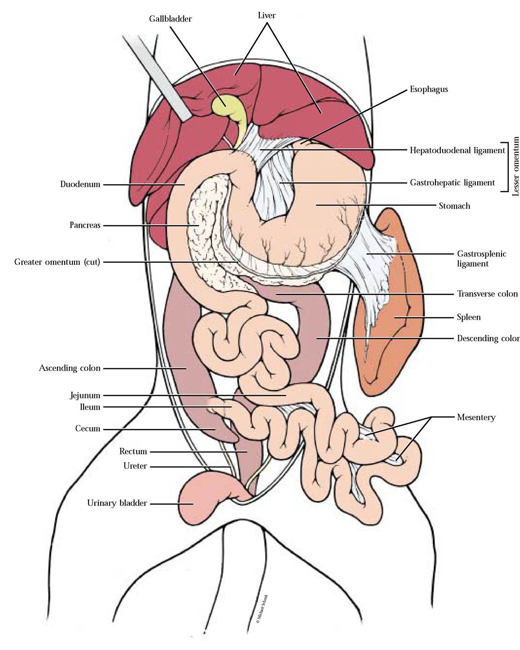
The stomach is a J-shaped organ lying mainly on the left side of the body. The convex left margin is known as the greater curvature and the concave right margin is known as the lesser curvature. Remember that the lesser omentum occurs between the liver and the lesser curvature of the stomach and the proximal end of the duodenum as the gastrohepatic and hepatoduodenal ligaments, respectively. From the greater curvature hangs the greater omentum. On the left side, the triangular gastrocolic ligament extends from the greater omentum to the mesocolon. That portion of the stomach below the cardiac sphincter is the cardiac region and the narrow portion connected to the intestine is the pyloric region. A muscular valve, the pyloric sphincter (pylorus), located at the distal end of the pyloric region regulates the movement of the stomach contents into the intestine. The large inflated portion between these two ends consists of the upper fundus and lower body [Figure 4-8].
Carefully make an incision along the greater curvature from the fundus to the pyloric area of the stomach making sure to avoid the greater omentum. If the stomach is full, carefully remove some of the material to observe the gastric rugae or folds in this organ that allow it to expand perceptively when food is eaten. If the stomach is full, the rugae may not be as obvious as in an empty stomach.
Small Intestine
From the stomach the contents of the alimentary canal move into the small intestine. In this region of the alimentary canal, most of the major chemical digestion of carbohydrates, proteins, and fats takes place. Some of the digestive enzymes are a structural part of the cells of the luminal epithelium of the small intestine while other essential enzymes and digestion facilitators are released from the pancreas and biliary system (liver and gallbladder) through ducts into the small intestine. This region is also another area where segmentation and peristalsis mix and move the contents along the alimentary tract. This is the major absorption site of nutrients and water in the digestive tract.
The lengthy small intestine actually occupies a minimal abdominal volume since it is greatly coiled. The small intestine is regionally subdivided into three areas, the duodenum, the jejunum, and the ileum [Figure 4-9]. Mesenteries play an important role in maintaining the position of the small intestine. The mesoduodenum supports the duodenum and head of the pancreas while the mesentery proper supports the jejunum and ileum. Observe the obvious lymph nodes associated with the mesentery proper. The triangular duodenocolic fold, on the right side of the body, anchors the duodenum to the mesocolon.
The very short proximal part of the small intestine about 12 to 18 cm (5-7 in) in length in an adult cat, known
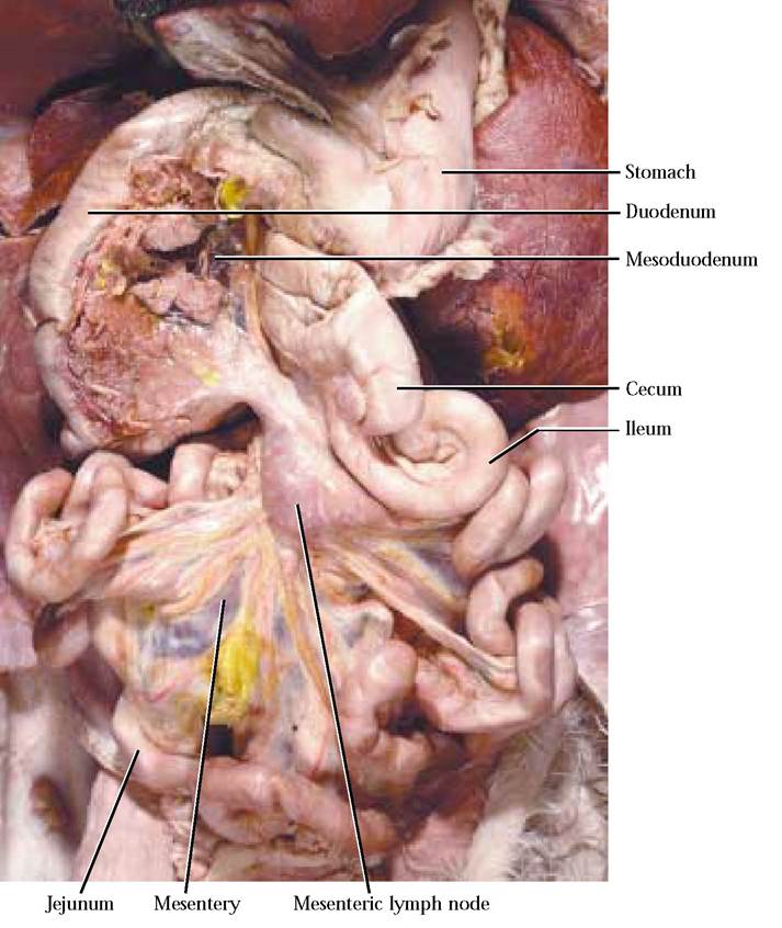
FIGURE 4-9 Regions of the small intestine.
Ileocecal valve Cecum
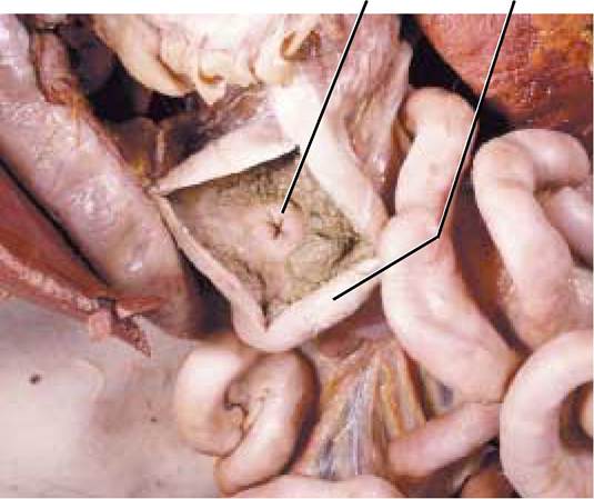
FIGURE 4-10 Ileocecal valve.
of bacteria. The vitamins are absorbed across the mucosa of the large intestine. The end result is the production of gases and semisolid feces that are eliminated from the body through the anus.
Anatomically, the proximal part of the large intestine is a blind diverticulum, the cecum. In humans, an elongate, fingerlike tube, the appendix, extends from the cecum. Cats do not possess an appendix.
From the cecum, the colon continues cranially on the right side of the body as the ascending colon, makes a left hand turn, crosses the abdominal cavity to the left side as the transverse colon where it curves caudally and continues as the descending colon terminating as the rectum [Figure 4-11]. The mesocolon suspends the large intestine from the parietal peritoneum of the dorsal wall. With the exception of the sigmoid colon in the human, this portion of the digestive tract is similar to that of the cat. The anus is the caudal opening of the digestive tract and is surrounded by sphincter muscles. A pair of scent glands, the anal glands, open into
as the duodenum, extends from the pylorus to the position of the duodenocolic fold. The distal-most portion of the small intestine is the ileum and the middle portion is the jejunum. At the junction of the ileum and the large intestine, on the righthand side of the body, is a doughnut-shaped muscular ileocecal valve, which regulates the movement of the contents of the small intestine into the large intestine as well as prevents the reflux of contents into the small intestine [Figure 4-10]. Carefully make a small incision with a sharp scalpel in the wall of the cecum at the site of the ileocecal valve and observe its shape [Figure 4-9]. You may have to clear any contents in this area to observe this valve.
Large Intestine
Further absorption of water occurs in this region along with fermentation, rotting of undigested material, and vitamin synthesis by resident colonies
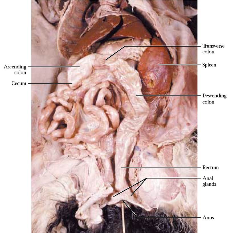
FIGURE 4-11 Regions of the large intestine.
the rectum near the anus [Figure 4-11]. Secretions are important in territorial marking and sexual attraction. Unfortunately, humans have no scent glands, but their alimentary canal is similar to the cat.
Accessory digestive organs
Liver
The largest internal organ in most mammals is the domeshaped liver that rests directly below the diaphragm. Among its many functions are chemical syntheses, detoxification, storage of metabolic products, bile production, etc. Actually, if you cannot pinpoint an organ where a particular function occurs, a good guess is the liver.
In the cat, the prominent, reddish-brown liver is divided into six lobes. A deep cleft from which the falciform ligament extends from the liver to the ventral body wall, separates the left and right halves. Identify a left medial and a left lateral lobe. Adjacent and to the right of the falciform ligament is the small quadrate lobe, which is partially united with the right medial lobe. The gallbladder is located in a semicircular depression, the cystic fossa, between the quadrate and right medial lobes. Just posterior to the right medial lobe is the right lateral lobe. The caudate lobe, just posterior to the right lateral lobe and sometimes appearing like a subdivision of that lobe, extends into the lesser omentum [Figure 4-12]. Observe the hepatorenal ligament extending between the caudate lobe and the right kidney.
The point at which the liver and the diaphragm actually contact one another is the site of the central tendon of the diaphragm [Figure 4-13]. Note that the falciform ligament continues as the coronary ligament to each side of the central tendon of the diaphragm. Also find the round ligament on the free surface of the falciform ligament. It represents the remnant of the umbilical vein of the cat during its fetal life when this vessel was essential in returning oxygenated blood from the placenta. The lesser omentum connects the liver to the lesser curvature of the stomach and to the duodenum.
An important digestive function of the liver is secretion of bile, a solution containing bile salts that are important in emulsifying fats, reducing the size of the fat globule, and increasing the surface area of the fat so that enzymatic digestion is facilitated. Additionally, bile is involved during fat absorption. Bile is secreted by liver cells and stored in the “bag” called the gallbladder. A number of ducts are associated with the liver and gallbladder. Many small hepatic ducts converge to form two or more quite prominent hepatic ducts leading from the left lobes and right lateral lobe of the liver that join the cystic duct, draining the gallbladder, forming the common bile duct [Figure 4- 14A and Figure 4-14B]. The common bile duct that extends parallel with the hepatic portal vein (appearing as a robust yellow
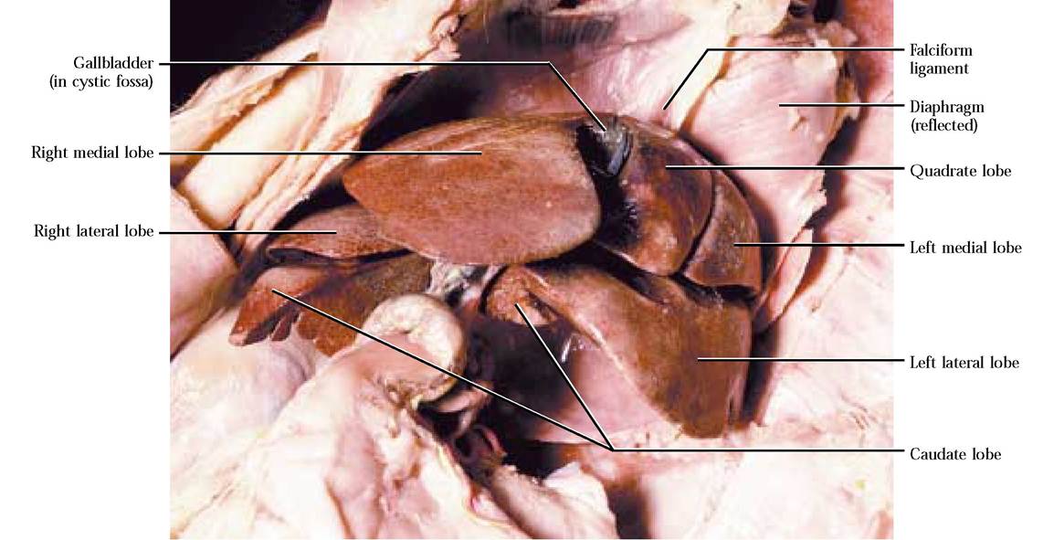
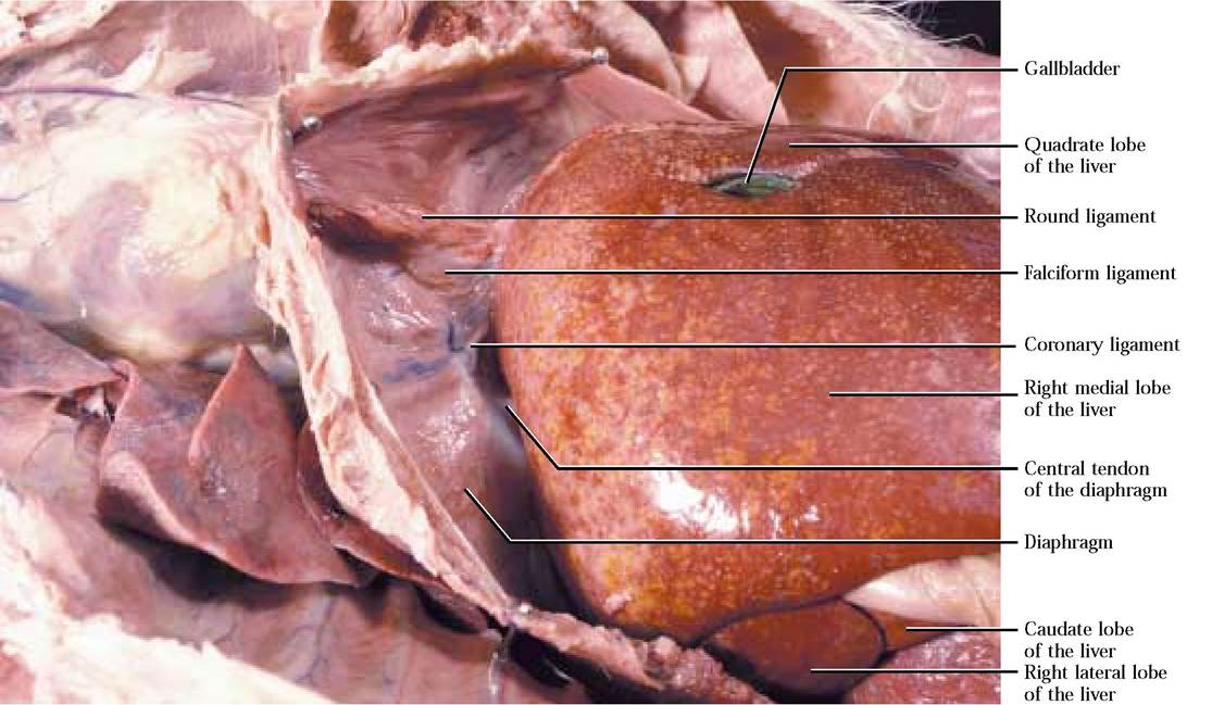
FIGURE 4-13 Relationship of the liver and diaphragm.
vessel in triply injected cats) and the relatively small hepatic artery (appearing as a red vessel) empties into the duodenum. All of these structures, with the exception of the hepatic ducts, accompanied by nerves and lymphatic vessels, form a tough tendinous right lateral border of the hepatoduodenal ligament of the lesser omentum.
To observe the duct system, carefully isolate with forceps, the common bile duct (a tannish or greenish, flat structure) within the border of the hepatoduodenal ligament beginning at its distal end where it joins the main pancreatic duct opening into the duodenum marked by a small pimplelike bump known as the hepatopancreatic ampulla (ampulla of Vater) [Figure 4-14A and Figure 4-14B]. The common bile duct is fragile and can easily be torn. From the ampulla it extends toward the liver where it is joined by the hepatic ducts from various lobes of the liver as well as the often greenish cystic duct leading from the gallbladder. You may have noticed that the gallbladder and surrounding liver tissue may be stained an olive green, the typical color of bile. Exposure of the hepatic ducts that have a tendency to shred and tear requires careful removal of hepatic tissue using forceps in a picking motion. The cystic duct is perhaps the easiest to find since it extends from the posterior end of the gallbladder. It is through this system of ducts that the bile is released under the influence of digestive tract hormones and fatty foodstuff in the chyme (intestinal contents).
In the human, there are only four lobes of the liver— the right and left separated by the falciform ligament, and a caudate and quadrate. The ductwork and functions are very similar.
Pancreas
This organ is actually one that is comprised of tissues derived from two different embryonic sources. Therefore, it should not be too surprising for you to learn that parts of it function as an endocrine gland producing essential hormones that control various metabolic activities throughout the body and the other portion produces essential digestive enzymes and a buffering solution of sodium bicarbonate, a cat's own antacid supply, actually only affecting the acidity of the small intestine rather than providing gastric relief.
The appearance of the pancreas is lobulated and glandular. It is the tannish, elongated organ whose head (duodenal portion) lies within the mesoduodenum and whose tail (gastrosplenic portion) lies within the dorsal part of the greater omentum near the greater curvature of the stomach [Figure 4-15]. Each part is drained by the main pancreatic duct that joins the common bile duct in the hepatopancreatic
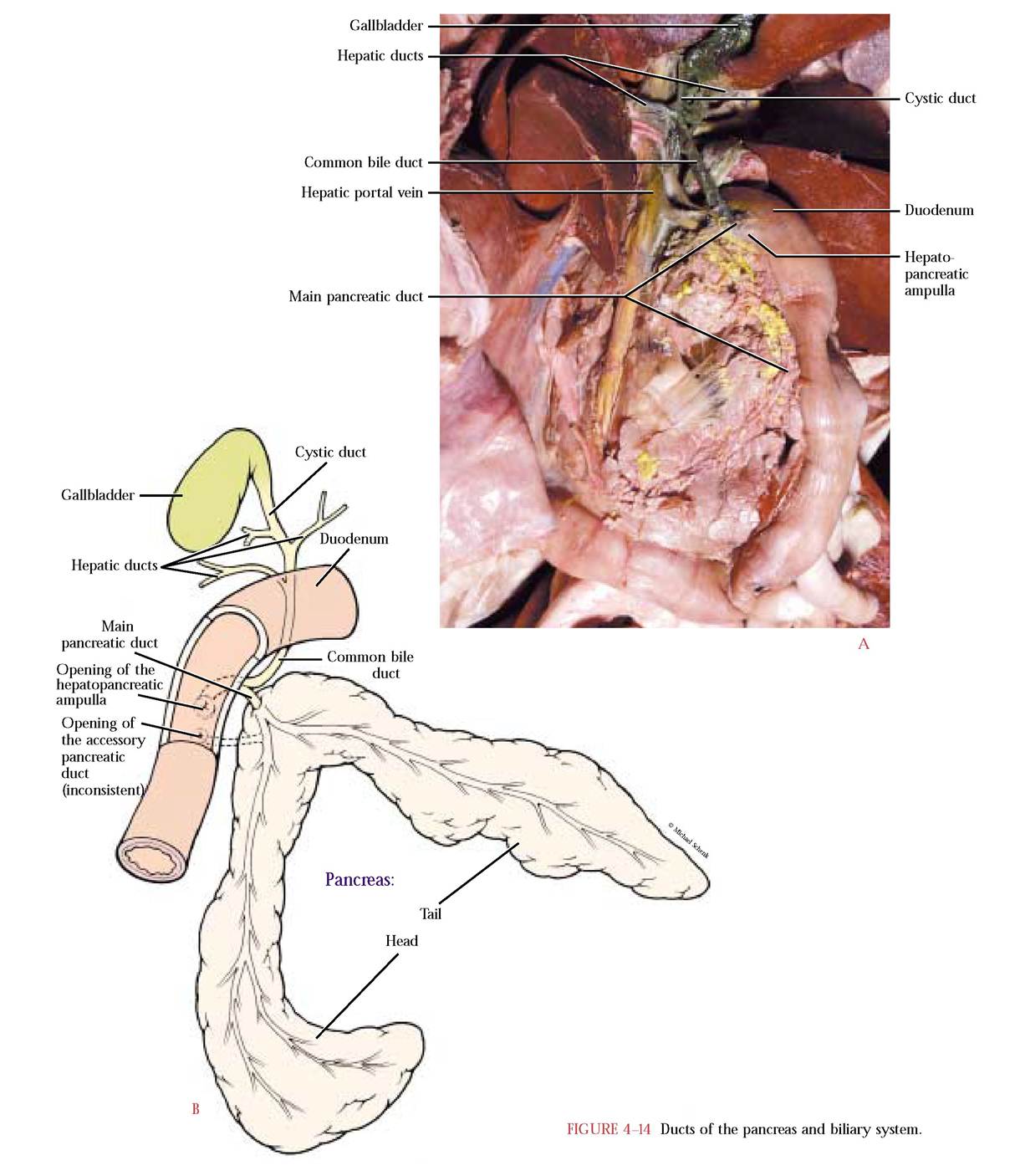
ampulla on the duodenum [Figure 4- 14A and Figure 4- 14B]. An accessory duct, not always present, may drain the gastrosplenic portion independently.
To observe the main duct, carefully and gently pick away pancreatic tissue along the duodenal border of the duodenal portion of the pancreas, starting from the ampulla and working toward the head end, taking special care to preserve all red-injected and yellow-injected blood vessels in the process. This duct is whitish with smaller branches feeding into it and is especially delicate. The accessory duct, leading from the gastrosplenic portion, quite often, is not as robust as the main duct and opens a short distance below the ampulla [Figure 4-14B]. This duct is often difficult to locate and may be absent in some cats. The pancreas and ducts in the human are very similar.
Spleen
The spleen is a large lymphoid organ often discussed with the digestive system because it occurs in the peritoneal cavity, along with the viscera. This large, tonguelike organ lies on the left side of the body and is anchored in the ventral layer of the greater omentum, the gastrosplenic ligament [Figure 3-6]. In humans the spleen is soft and fist-sized.
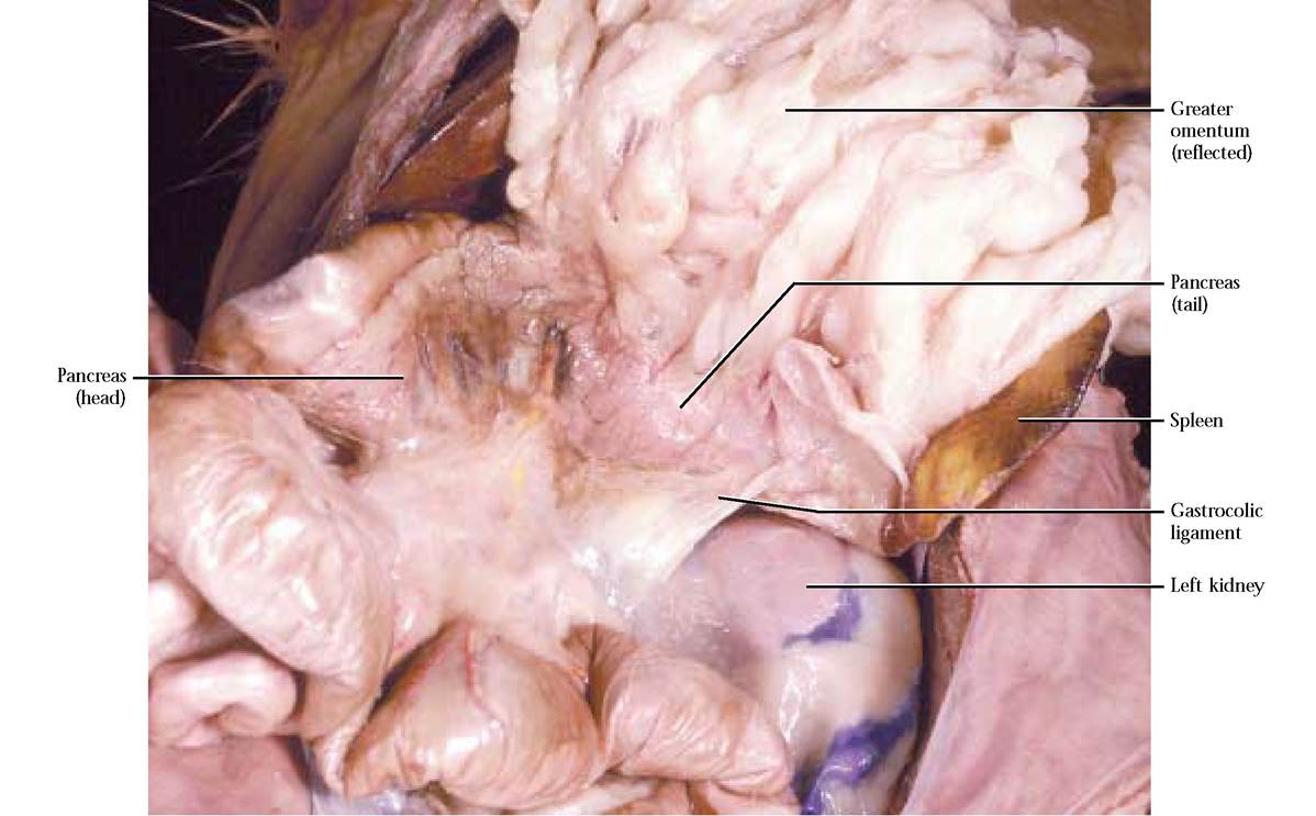
FIGURE 4-15 Pancreas.