Respiratory System
Gas exchange of oxygen and carbon dioxide is a life sustaining function of the respiratory system. Among most adult tetrapod vertebrates, cats and humans included, the organs facilitating this exchange are the lungs.
They are located internally and generally found within the thoracic cavity of the body. The process of gas exchange, diffusion, occurs across the moist, inner surfaces of the lungs into capillaries. The spongy lungs are made up of great numbers of gas-filled small spheres or alveoli where this diffusion takes place. The vast number of alveoli makes it possible to maximize the surface area in a reasonably sized organ such as the lung.A system of tubes, the trachea, various bronchi and bronchioles conveys the oxygen-rich air from the atmosphere to the lungs and the oxygen-poor air back to the atmosphere [Figure 5-1]. The diameter of these tubes decreases while the number increases as they extend from the nasal cavity to the alveoli. The overall effect is to increase the respiratory surface area dramatically.
Among some reptiles and all mammals the palate described in the digestive system separates the oral
and the nasal cavities. In the digestive system of mammals, the palate serves as the roof of the oral cavity and as the floor of the nasal cavity of the respiratory system. This “shelf” allows a mammal to chew and hold food items in the mouth and breathe at the same time. It also allows mammals to smell and taste food for this reason.
Because some organs are common to the digestive and respiratory systems, we suggest that the digestive system be dissected first.
OPENINGS AND SPACES
The pathway by which air moves from the atmosphere to the alveoli begins through the nostrils or external nares and into the nasal cavities.
In these cavities the air is warmed, moistened, filtered, and smelled. From the cavities it passes through the internal nares, or choanae, and into the nasopharynx, one part of a more extensive region called simply the pharynx, a space shared with the digestive system. The nasopharynx extends from the internal nares to the free edge of the soft palate. The air then moves into the laryngopharynx. From this space, the air moves into the larynx [Figure 4-5].| LARYNX, TRACHEA, AND BRONCHI |
Before the larynx is dissected, locate the paired thyroid glands situated on either side of the trachea, just caudal to the larynx [Figure 5-2]. In particular, be aware of the isthmus, a narrow band of tissue that extends at various angles between the two lobes.
The larynx is cartilaginous and consists of several single and paired elements. One of the single elements is the epiglottic cartilage that strengthens the epiglottis. The function of the epiglottis is to prevent the contents of the oral cavity from entering the trachea during swallowing. As the material is swallowed, the larynx is pulled cranially by muscles, causing the epiglottis to block off the glottis, a slit between the vocal cords. Air passes into the larynx through this slit. To view the epiglottis in a bisected specimen, see Figure 4-5. With a specimen dissected into a dorsal and ventral half, pull the tongue forward while peering into the pharyngeal region and observe the epiglottis, see Figure 4-6.
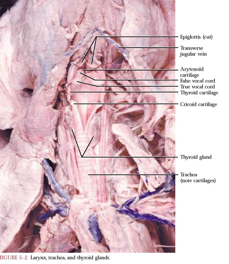
A second, very large single cartilage noticeable ventrally is the thyroid cartilage [Figure 5-2 and Figure 4-5]. Observe that this cartilage is continuous dorsally. To see this structure better, you should remove the musculature obscuring it. In humans, the thyroid cartilage is commonly called the Adam's apple and exhibits a distinct sexual dimorphism, i.e., in males it is much more prominent.
Caudal to the thyroid cartilage is the ring-shaped cricoid cartilage [Figure 5-2 and Figure 4-5]. The dorsal wall of the larynx consists primarily of the broad part of this cartilage. You may also want to clear musculature from this cartilage.
Small, paired cartilages, the arytenoid cartilages [Figure 5-2], sit on the dorsal rim of the cricoid cartilage and abut the dorsal projections of the thyroid cartilage. Some other inconspicuous cartilages are part of the larynx, but are difficult to see. In humans, these cartilages, the cuneiform and the corniculate, are more easily identified, in addition to the cartilages described in the cat.
With a sharp scalpel, make an incision in the midventral wall of the larynx, perhaps extending it into the trachea caudally. With scissors, continue the cut cranially, cutting the epiglottis. These directions apply only to the dorsoventral head dissection. Carefully reflect the walls of the larynx. Observe two pairs of tissue folds at the cranial end of the larynx. The cranial folds, the false vocal cords, often appearing tannish, extend from the arytenoid cartilages to the epiglottis. The caudal folds, the true vocal cords, appearing as whitish bands, extend from the arytenoid cartilages to the thyroid cartilage [Figure 5-2 and Figure 4-5]. Vocalization in cats is the result of air movement across the true vocal cords, while purring seems to be related to the vibration of the vocal cords during air movement through the glottis. In humans, the false vocal cords are not involved in sound production.
The trachea extends caudally from the larynx to the root of the lungs [Figure 5-2 and Figure 4-5]. This large diameter respiratory tube is the main passageway for air from the larynx to the lungs. Observe the C-shaped cartilages that reinforce the wall of the trachea, with the dorsal open space of the “C” reinforced with muscle. This anatomical configuration allows compression during the act of swallowing but prevents collapse during respiration.
The lobulated, glandular tissue, the thymus, lying along the ventral aspect of the trachea and heart should be conserved. This important endocrine gland varies in size with the age of the animal, also true in human beings.Division of this tube within the tissue of the lungs occurs with a resulting interconnected network of air conducting tubes often referred to as a bronchial or respiratory tree. The first division of the trachea forms the primary bronchi. These are subdivided sequentially into secondary and tertiary bronchi [Figure 5-3]. Further branching results
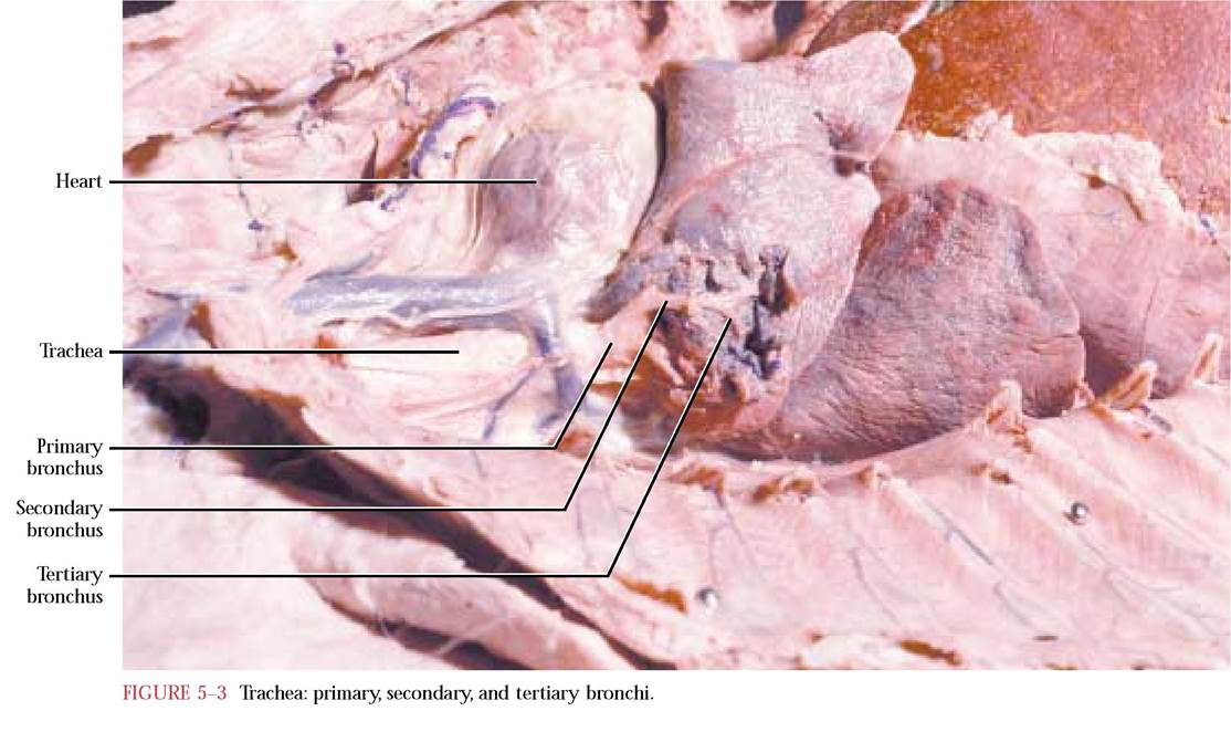
in bronchioles and alveolar ducts which terminate in alveoli, illustrating the decreasing diameter but increasing surface area mentioned previously. The supporting cartilage of these tubes decreases proportionately to the increased surface area and elasticity of the tree.
To observe the primary bronchi, reflect the lungs medially and carefully pick away lung tissue at the level of the root of the lungs. Be careful to avoid the pulmonary vessels in this area that enter and leave the lung at the hilus. The bronchi appear as whitish, shiny tubes containing cartilage. Since the rest of the tree is enclosed within the lungs, we do not recommend further dissection. Your instructor may wish to provide a demonstration dissection.
LUNGS
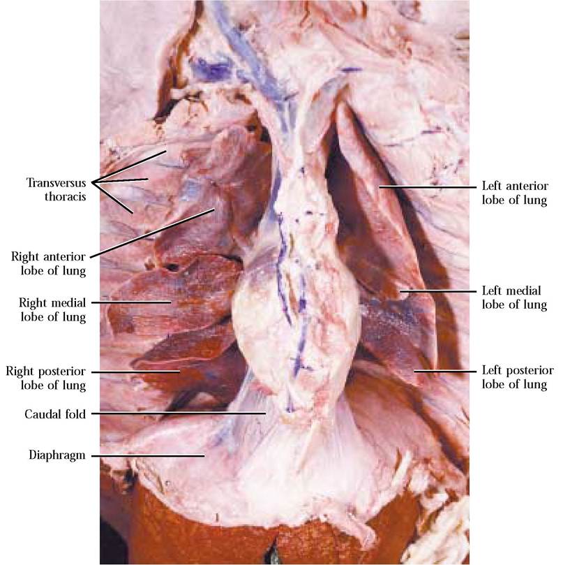
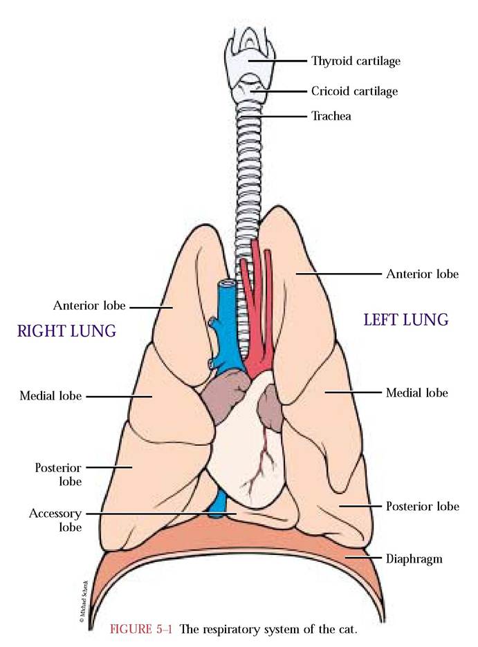
The organs known as lungs are spongy, primarily due to the terminal structures of the respiratory tree called alveoli. Air moves through the tubular network into the alveoli, small,
DIAPHRAGM
The often mentioned muscular partition between the thoracic and abdominopelvic cavities is the diaphragm [Figure 5-4]. Mammals are the only vertebrates in which is found a complete muscular separation of these two cavities. Through this partition passes the esophagus, the posterior vena cava, and the aorta.
This organ is an essential component involved in the process of respiration.
When this dome-shaped muscular partition contracts, moves caudally and flattens, the volume of the pleural cavities increases, with a concomitant decrease of pressure, and air moves into the respiratory tract. In contrast, when the diaphragm relaxes and returns to its domed position, the volume of the pleural cavities decreases and the pressure increases with the result that air moves into the atmosphere, according to the laws of gas physics.thin terminal air sacs, where gas exchange occurs. The left lung is subdivided into three lobes, the anterior, medial, and posterior. The right lung is also similarly divided with the exception of the posterior that is further subdivided forming an accessory lobe. Remember that this lobe of the lung projects into a mesentery pocket called the caval fold, while its left hand mesentery is the mediastinal septum [Figure 5-4]. In addition, remember that the lungs are suspended within the pleural spaces by the pulmonary ligaments [Figure 3-4B]. In human beings, a similar pattern of organ construction occurs with the exception of the number of lobes of the lungs. In humans, the left lung is subdivided into two lobes while the right has three.