Each Heartbeat Consists OfVentricuIar Systole and Ventricular Diastole
The heart is actually two pumps (two ventricles) that work together, side by side. Each ventricular pump works in a cycle, first relaxing and filling with blood and then contracting and ejecting some blood.
In each cardiac cycle (heartbeat) the left ventricle takes in a volume of blood from the pulmonary veins and left atrium, then ejects it into the aorta. The right ventricle takes in a similar volume of blood from the systemic veins and right atrium, then ejects it into the pulmonary artery.Figure 21-1 shows the events of a single cardiac cycle. A normal electrocardiogram (ECG) tracing is presented at the top of the figure. Atrial contraction is initiated by atrial depolarization, which is indicated by the P wave. Ventricular contraction is initiated by ventricular depolarization, which is indicated by the QRS complex. The period of ventricular contraction is called ventricular systole. Blood is ejected from the ventricles during ventricular systole. Each systole is followed by ventricular diastole, during which the ventricles relax and refill with blood before the next ventricular systole. Note that ventricular diastole corresponds to the period
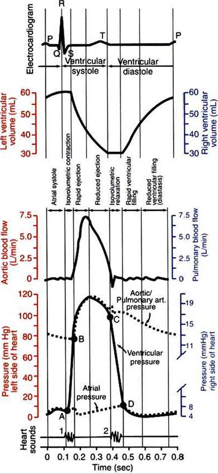
FIGURE 21-1 Events and terminology associated with one cardiac cycle (heartbeat) in a normal dog. Vertical scales on the left side of the graphs (red} are for the left side of the heart. Vertical scales on the right side of the graphs (blue} are for the right side of the heart. In the graph of ventricular pressure, point A indicates closure of the mitral and tricuspid valves (the atrioventricular valves); point B indicates opening of the aortic and pulmonic valves; point C indicates closure of the aortic and pulmonic valves; and point D indicates opening of the mitral and tricuspid valves. See text for details.
between a T wave and the subsequent QRS complex, when ventricular cells are at resting membrane potential.
The ventricles do not empty completely during systole. As shown in the graph of Ventricularvolume (Figure 2l-1, second from top), each ventricle of a large dog contains about 60 mL of blood at the end of diastole. This is called end-diastolic volume. During systole, about 30 mL of this blood is ejected from each ventricle, but 30 mL remains. This is called end- systolic volume. The volume of blood ejected from one ventricle in one beat is called stroke volume, expressed as follows:
Stroke volume = End-diastolic volume - End-systolic volume The fraction of end-diastolic volume that is ejected during ventricular systole is called the ejection fraction, as follows:

In the example depicted in Figure 21-I, ejection fraction is 50%. Values between 50% and 65% are typical for resting dogs.
As shown in Figure 21-1, left ventricular pressure is low at the beginning of ventricular systole, but the powerful contraction of the ventricular muscle causes the ventricular pressure to increase rapidly. The increase in left ventricular pressure causes a momentary backflow of blood from the left ventricle to the left atrium, which closes the left atrioventricular (AV) valve (the mitral valve). Blood is not immediately ejected from the left ventricle into the aorta at the beginning of systole, because the aortic valve remains closed until the left ventricular pressure exceeds the aortic pressure. Therefore, ventricular volume remains unchanged during this first phase of systole, which is aptly named isovolumetric contraction.
When left ventricular pressure does rise above aortic pressure, the aortic valve is pushed open, and there is a rapid ejection of blood into the aorta. Rapid ejection is followed by a phase of reduced ejection of blood as both ventricular pressure and aortic pressure pass their peak (systolic) values and begin to decrease.
(During the period of reduced ejection, the ventricular pressure actually falls below the aortic pressure, but ejection continues for a few moments, because the blood flowing out of the ventricle is carried along by the momentum imparted to it during rapid ejection.) As the ventricular pressure continues to decrease, ejection comes to an end. A momentary backflow of blood from the aorta into the left ventricle closes the aortic valve. The closure of the aortic valve demarcates the end of ventricular systole and the beginning of ventricular diastole.During the first phase of ventricular diastole, the ventricular muscle relaxes, and left ventricular pressure declines from a value near aortic pressure to a value near left atrial pressure. However, no filling of the ventricle can occur because the mitral valve remains closed until left ventricular pressure drops below left atrial pressure. This first phase of ventricular diastole is called isovolumetric relaxation because there is neither filling nor emptying of the ventricle.
When left ventricular pressure does fall below left atrial pressure, the mitral valve is pushed open, and ventricular filling commences. First, there is a period of rapid ventricular filling, which is followed by a phase of reduced ventricular filling (diastasis). Diastasis persists until the sinoatrial node cells initiate an atrial action potential and atrial contraction (atrial systole). In a resting dog, as depicted in Figure 21-1, ventricular volume is nearly at its end-diastolic level even before atrial systole. Typically, 80% to 90% of ventricular filling occurs before atrial systole. Atrial systole simply “tops up” the almost-full ventricles. An important clinical consequence of this fact is that the heart in a resting animal pumps a nearly normal cardiac output even in the absence of properly timed atrial contractions (e.g., during atrial fibrillation). During exercise, however, atrial contractions make a relatively greater contribution to ventricular filling because the rapid heart rate in exercise leaves a shorter time for diastolic filling.
Therefore, animals with atrial fibrillation typically exhibit exercise intolerance. Ventricular filling also becomes more dependent on atrial systole in patients with certain valve defects, such as narrowing of the mitral valve (mitral stenosis).At the end of atrial systole, the atria begin to relax. The left atrial pressure drops slightly. The ventricles then begin to contract, and there is a momentary backflow of blood from the left ventricle to the left atrium. The mitral valve closes, which marks the end of ventricular diastole and the beginning of another left ventricular systole.
By definition, the cardiac cycle is divided into ventricular systole and ventricular diastole. Closure of the mitral valve marks the beginning of ventricular systole. Closure of the aortic valve marks the beginning of ventricular diastole. Atrial systole takes place during ventricular diastole.
The preceding six paragraphs discussed pressure changes in the left atrium, left ventricle, and aorta. However, all the events of the cardiac cycle also take place on the right side of the heart. Therefore, all the statements made about the left side of the heart also hold true for the right side of the heart; simply substitute “pulmonary artery” for “aorta,” “pulmonic valve” for “aortic valve,” and “tricuspid valve” for “mitral valve.” As indicated in Figure 21-1, the ventricular volumes are similar for the left and right sides, and so are the blood flow rates. The pressures, however, differ greatly on the two sides. Systolic (peak) pressure in the right ventricle and pulmonary artery is only about 20 mm Hg, whereas systolic pressure on the left side of the heart reaches 120 mm Hg. This explains why there are different scales on the pressure axes in Figure 21-1 for the left and right sides of the heart.
The timing of the two major heart sounds is also shown in Figure 21-1 (bottom). The first heart sound is associated with the closure of the AV valves (the mitral and tricuspid valves).
The actual closure of the valves does not make this sound; the valve leaflets are so light and thin that their closing would be almost silent. However, there is a momentary backflow of blood from the ventricles to the atria at the beginning of ventricular systole. When this backflow of blood is brought to a sudden stop against the closing valves, momentary vibrations are created in the blood and in the cardiac walls. These vibrations create the heart sound.The second heart sound is associated with closure of the aortic valve on the left side of the heart and the pulmonic valve on the right side of the heart. It is usually briefer, sharper, and higher pitched than the first heart sound. Again, what makes the sound is not the valve leaflets closing, but rather the reverberation produced when the momentary backflow of blood into the ventricles is brought to a sudden stop by closure of the valves. The closures of the aortic and pulmonic valves are normally simultaneous. Under certain circumstances, however, the two valves close at slightly different times, and the second heart sound is heard as two distinct sounds in quick succession; this condition is called a split second heart sound.
The AV valves close at the beginning of ventricular systole, and the aortic and pulmonic valves close at the end of ventricular systole. Therefore, ventricular systole is sometimes defined as the part of the cardiac cycle between the first heart sound and the second heart sound.