Embryology
The nervous system is the first organ system to begin to form in the embryo (see Chapter 3). Shortly after gastrulation, ectodermal cells on the dorsum just cranial to the primitive streak begin to proliferate and differentiate into a neural plate.
The neural plate proliferates faster along its lateral margins than on the midline, creating the neural groove, the edges of which (the neural folds) ultimately meet dorsally to form the neural tube (Fig. 9-5). The entire CNS is formed from the cells of the neural tube. The lumen of the neural tube persists in the adult as the central canal of the spinal cord and as the ventricles of the brain (discussed later).Closure of the neural tube is not simultaneous throughout the embryo. Fusion develops first at the level that will eventually become the medulla oblongata (the most caudal part of the brainstem) and proceeds craniad and caudad from there. The openings at the cranial and caudal end of the closing tube are called the rostral and caudal neuropores, respectively (see Fig. 3-4).
The rostral neuropore closes early in development; failure to do so disrupts development of the brain, leading to profound underdevelopment of the head. In its most severe form (short of embryonic death), anencephaly (a complete absence of the
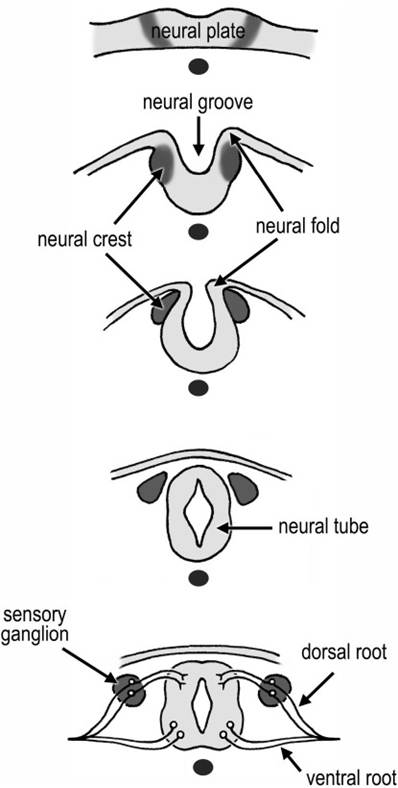
Figure 9-5. Formation of the neural tube. The thickened ectoderm of the neural plate develops into a groove that subsequently fuses on the dorsal side to form a closed tube. Neural crest cells adjacent to the neural folds differentiate into many tissues, including the neurons of the ganglia.
cerebrum, often with concurrent absence of meninges and skull) results. The caudal neuropore closes later. Failure of closure in the caudal part of the neural tube results in a variety of spinal cord abnormalities called myelodysplasias.
These are sometimes also associated with vertebral anomalies, such as spina bifida.As the edges of the deepening neural groove approach one another at the dorsal midline, a longitudinal column of cells differentiates at the union between the ectoderm and the neuroectoderm on each side of the groove. These cells, the neural crest, end up lateral to the neural tube on each side of it and eventually form sensory and autonomic ganglion cells, schwann cells, and other related tissues. in addition, the neural crest gives rise to a variety of other cell types, including parts of the meninges and many of the bones and muscles of the head.
Development of the spinal cord continues by an increase in the thickness of the wall of the neural tube. As cells divide and differentiate, three concentric layers of the neural tube emerge: an inner ventricular zone, a middle intermediate zone, and a superficial marginal zone (Fig. 9-6).
The thin ventricular zone of cells (also called ependymal or germinal zone) surrounds the lumen of the neural tube and is the site of mitosis of neuronal and glial precursors in the developing nervous system. it will ultimately form the ependyma of the central canal of the spinal cord and of the ventricles of the brain.
As cells are born in the germinal layer, they migrate outward to form the intermediate zone (also called mantle zone). The intermediate zone comprises neurons and neuroglia and becomes the gray matter near the center of the cord. The dorsal parts of the intermediate zone develop into the dorsal horns. It is here that sensory processing takes place. The ventral intermediate zone becomes the ventral horns, the location of the motor neurons whose axons will extend out into the periphery to innervate muscles and glands.
The marginal zone, which is most superficial, consists of nerve processes that make up the white matter of the spinal cord. The white color comes from the fatty myelin sheaths. The spinal cord white matter is divided into dorsal, lateral, and ventral funiculi, which are delimited by the dorsal and ventral horns of gray matter.
Development of the brain (Fig. 9-7) begins before the neural tube is fully closed caudally. it grows rapidly throughout embryonic and
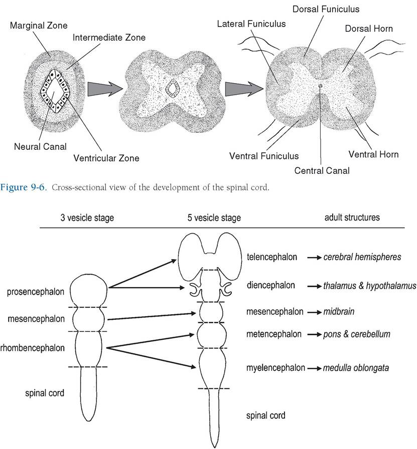
Figure 9-7. Dorsal view of the neural tube. The early brain divides into three vesicles that further differentiate into five vesicles. These give rise to the main regions of the adult brain. Notice the optic cups (primitive retinae and optic nerves) developing from the diencephalon.
fetal life and into the neonatal period. The first gross subdivisions of the brain create the three- vesicle stage. These subdivisions, which consist of three dilations of the presumptive brain, are the prosencephalon, or forebrain; mesencephalon, or midbrain; and rhombencephalon, or hindbrain. The prosencephalon develops lateral extensions, the optic vesicles, the precursors of the optic nerves and retinas.
With further development, the three vesicles differentiate into five distinct regions. In this five-vesicle stage of development, the prosencephalon further subdivides to form the telencephalon (future cerebrum) and the diencephalon, and the rhombencephalon divides into the metencephalon (future pons and cerebellum) and the myelencephalon (future medulla oblongata). The mesencephalon does not subdivide; it will become the future midbrain (often persistently called the mesencephalon).