Microscopic Neuroanatomy
The individual nerve cell is called a neuron (Fig. 9-2). Neurons possess the usual features of cells, but in keeping with their function of communication over long distances, they also exhibit a number of specializations.
Each neuronal cell body gives rise to one or more nerve processes, cytoplasmic extensions of the cell. The nerve processes are called dendrites if they transmit electrical signals toward the cell bodies; they are called axons if they conduct electrical signals away from the cell bodies. The axon (each neuron gives rise to only one, which usually branches) arises from a conical mound of cytoplasm, the axon hillock, and its terminus branches into an arborization called the telo- dendrion. The telodendrion makes contact with other neurons or effector organs (targets). In mammals, effector organs stimulated either directly or indirectly by the telodendrion include muscle and glands.The junction between the axon of one neuron with another neuron or target cell is the synapse. The neuron belonging to the axon is the presynaptic neuron, and the one receiving information from the axon is the postsynaptic neuron. A synapse may be between the axon of one neuron and the cell body, dendrites, and/or axon of the postsynaptic neuron. Typically, each neuron synapses with many other neurons through the extensive branching of its terminal ends and of its axon; branches of the main axon are axon collaterals.
Neurons may be classified morphologically according to their number of nerve processes (Fig. 9-3). Unipolar neurons have one process; true unipolar neurons are seen only during development. Bipolar neurons have one dendrite and one axon; these are common in sensory systems. Many sensory neurons have their single dendrite and axon fused so as to give the appearance of a single process; this configuration is pseudounipolar. Multipolar neurons have a number of dendrites in addition to their single axon.
Most neurons are multipolar in nature.Nervous tissue consists of not only neurons but also supportive cells. Within the CNS, these supportive cells are the neuroglia, comprising a variety of glial cells, whereas most of the supporting tissue of the peripheral nerves is ordinary white fibrous connective tissue.
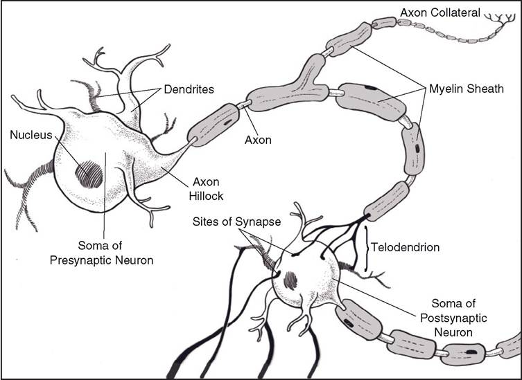
Figure 9-2. Cellular anatomy of a multipolar neuron.
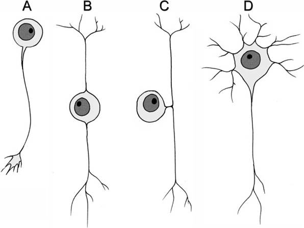
Figure 9-3. Morphological types of neurons. Classification is based on number of processes extending from the cell body. Unipolar neurons (A) are not found in adult vertebrate nervous systems; the unipolar neuron here is depicted with a growth cone at the tip of its axon, illustrating its developmental nature. Bipolar (B) and pseudounipolar (C) neurons are characteristic of sensory systems. Most neurons in the nervous system are multipolar (D) and can assume a variety of shapes.
Nerve fibers may be myelinated or unmyelinated. Myelinated fibers are surrounded by a white sheath of fatty material, or myelin. The myelin sheath actually consists of many layers of cell membrane of a specialized glial cell wrapped around axons so that in cross-section the myelin sheath resembles a slice of jelly roll. In the PNS, the myelinating cell is the Schwann cell (neurolemmocyte), whereas in the CNS, the oligodendrocyte fulfills this function. unmyelinated nerve fibers are not exposed directly to the extracellular fluid; rather, they are simply invaginated into the cell membrane of an adjacent glial cell. Axons covered in this
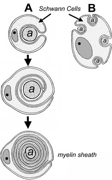
Figure 9-4. A) Cross-section of the development of a myelinated axon. In the peripheral nervous system, the myelinating glial cell is a Schwann cell; in the CNS, the oligodendrocyte lays down myelin wraps. B) Some Schwann cells in the peripheral nerves envelop multiple axons without forming the wrappings of myelin. Axons thus embedded are considered nonmyelinated.
way are not myelinated, which is very specifically the condition of being wound in multiple layers of glial cell membrane. Several unmyelinated fibers may be invaginated into separate areas of the same Schwann cell (Fig. 9-4).
Groups of nerve cell bodies within the CNS are generally called nuclei, while groups of nerve cell bodies in the PNS are called ganglia. Do not confuse a nucleus of the CNS with the nucleus of an individual cell. Bundles of nerve processes within the CNS are frequently called tracts, or fasciculi, and bundles of processes in the PNs are called nerves. In general terms, aggregates of neuronal cell bodies form the gray matter of the CNS, whereas regions characterized primarily by tracts are white matter.