FUNCTIONS OF DNA AND RNA
1. What comprises each chromosome?
2. What are the chemical bases that make up the two nucleotide chains of DNA?
3. How are the two nucleotide chains bound together and what are the complementary positions of the bases?
4.
What is the relationship of the histone proteins to the nucleotide chains?5. Where is the chemical location for the beginning of DNA.replication?
6. What is the point of attachment of the two newly formed chromosomes (chromatids) called?
7. Describe a gene as related to the DNA molecule.
8. What are the four stages of mitosis?
9. What is the name of each pair of replicated centrioles?
0. Visualize and describe each of the four stages of mitosis, recognizing interphase as a period between successive sequences.
11. Is DNA in the nucleus able to enter the cytoplasm to initiate the synthesis of protein?
2. What are the separate functions of mRNA, tRNA, and rRNA?
3. How is protein synthesis related to allergies and tissue rejection by individual animals?
DNA and Its Replication
The nucleus is composed mostly of the chromosomes, those structures providing for inherited and individual characteristics of an animal. Each chromosome is made up of a large molecule of DNA wrapped in the form of double helices (a helix is a spiral form) around a core of histone proteins. DNA is made up of two extremely long polynucleotide chains, each containing the purine bases adenine and guanine and the pyrimidine bases thymine and cytosine (Figure 1-3). A nucleotide is formed by the combination of one molecule of phosphoric acid, one molecule of deoxyribose, and one of the four bases. The chains are bound together by hydrogen bonding between the bases, with adenine bonding to thymine and guanine to cytosine. The bonding relationship is referred to as complementary (i.e., they are not identical). Whenever adenine appears on one strand, thymine will be in the same position on the opposite strand.
The histones are positively charged proteins that associate strongly with DNA by ionic interactions with its many negatively charged phosphate groups. About half of the mass of chromatin is DNA and half is histones. The whole complex of DNA and histones is called chromatin. Before cell division, the coiling around the histone proteins is loosened and replication of DNA begins by splitting the double helices at the point of junction of complementary bases. The separate strands then serve as a template for the formation of its complementary base when replication (making a facsimile or copy) takes place (Figure 1-4). The result is that each of the two original strands of each chromosome is now paired with a new complementary strand, forming two spiral helix chromosomes wherever there was one before. The two newly formed chromosomes remain temporarily attached to each other (until the time for mitosis) at a point called the centromere, located near their center. These duplicated but attached chromosomes are called chromatids. The units of heredity are the genes on the chromosomes, and each gene is a portion of the DNA molecule. Large numbers are attached end-on-end on the long, double-stranded, helical molecules of DNA that have molecular weights measured in the billions.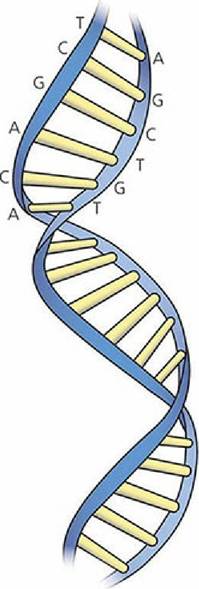
■ FIGURE 1-3 Two polynucleotide chains constitute the double helix of the DNA molecule. Obligatory base pairing occurs between A (adenine) and T (thymine), and also between G (guanine) and C (cytosine). The chains are held together by hydrogen bonding between bases. Histone proteins form a core between the nucleotide chains.
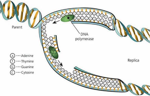
■ FIGURE 1-4 Replication of DNA. Coiling around histone proteins is loosened and the double helices split at a point of junction of complementary bases. The separate strands serve as a template for formation of its complementary base.
Two new double-helix chromosomes formed where only one was before. (From Frandson RD, Wilke WL, Fails AD. Anatomy and Physiology of Farm Animals. 7th edn. Ames, IA: Wiley-Blackwell, 2009.)Mitosis
Mitosis is the division of somatic cells (body cells, as opposed to reproductive cells) in which complex nuclear division precedes cytoplasmic fission and involves a sequence of four stages: prophase, metaphase, anaphase, and telophase (Figure 1-5). The period between successive sequences is called interphase. Prior to initiating mitosis two important things must be duplicated: (1) The chromosomes (DNA) and (2) the centrosome. The centrosome is made up of two centrioles and when duplicated each will serve as a future pole during cell division. In Figure 1-5A the centrosome has duplicated, as indicated by the four centrioles located within it. As prophase begins, the two centrosomes separate and begin to move to opposite poles of the cell (Figure 1-5B). In the process, each centrosome sends microtubules in all directions and is now referred to as an aster. When microtubules from each aster connect with each other they form the mitotic spindle. As the cell enters metaphase (Figure 1-5C) the nucleus breaks down and allows some of the spindle microtubules to interact with a specialized region on the duplicated chromosomes, referred to as the kinetochore. The microtubules that connect with the kinetochore are referred to as kinetochore microtubules while those that connect the two poles are interpolar microtubules. The interaction between the microtubules and chromosomes results in the chromosomes being aligned at the midway point between the spindle poles. As anaphase starts, the duplicated chromosomes separate and are pulled in opposite directions towards one of the spindle poles by the kinetochore microtubules (Figure 1-5D). In telophase, the chromosomes arrive at their respective spindle pole and a new nucleus forms around the chromosomes (Figure 1-5E). At the end of anaphase/beginning of telophase, a constriction called the cleavage furrow develops around the middle of the cell, which will finish by separating the cytoplasm so two daughter cells are created, each with their own nucleus and cytoplasm.
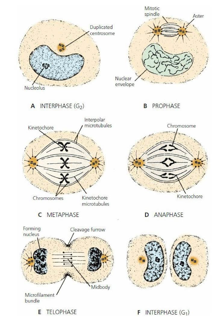
■ FIGURE 1-5 Diagrammatic representation of the stages of mitosis. See text for details. (From Cormack DH. Ham’s Histology. 9th edn. Philadelphia, PA: JB Lippincott Company, 1987.)
RNA and Protein Synthesis
Genes control the formation of cell proteins by a complex process of coding, the so-called genetic code. Because of its large size and inability to enter the cytoplasm, DNA in the nucleus is not able to directly control the synthesis of protein that occurs in the cytoplasm. RNA molecules that are synthesized from DNA accomplish this. The first of these, messenger RNA (mRNA), moves into the cytoplasm through nuclear pores carrying the code for the synthesis of proteins (transcription) and establishes a position with a granular ER ribosome where protein molecules are made. A second, transfer RNA (tRNA), is synthesized by DNA and moves to the cytoplasm, where it picks up an amino acid and carries it to the mRNA. There the amino acid is fitted into the code for the production of a specific protein molecule (translation). Each of 20 tRNAs are specific for each of the 20 amino acids. The third type of RNA is ribosomal RNA (rRNA), found in ribosomes. It is believed that it serves as a physical structure on which the protein is formed. The sequence of protein synthesis is shown in Figure 1-6. Because of the transfer of information required for protein synthesis from DNA molecules in the nucleus, it can be seen that proteins are specific to each individual animal. Introduction of proteins foreign to an animal results in allergies, tissue rejection, and other incompatibilities.
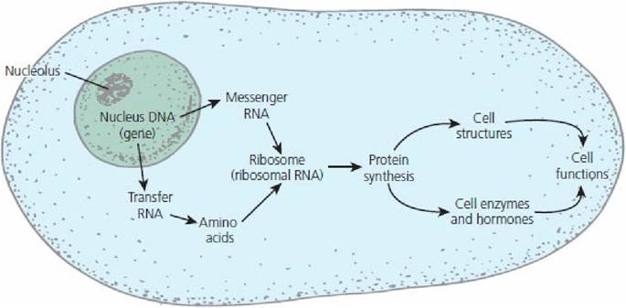
■ FIGURE 1-6 A schematic summary of genetic coding and its role in protein synthesis and related cell functions.
■