Endocrine System
Although the organs in this chapter are collectively known as a system, they are not physically connected in many cases. The functions of these organs, often known as glands, are exquisitely coordinated when they are operating normally.
When they are not, often the result is a dysfunctional organism, cat or human. Secretions of these ductless glands, called hormones, act as regulatory chemicals, often interacting among themselves to control a complex series of cellular activities. The hormones are transported in the blood of the circulatory system to their target tissues where they are attracted to very specific receptor sites on the surface of these cells, mediating their characteristic activities.In this chapter, individual endocrine glands will be identified with their position and function.
Pituitary gland
OR HYPOPHYSIS
The pituitary gland is connected to the ventral surface of the hypothalamus of the brain by a stalk (the infundibulum) and rests in the sella turcica of the sphenoid [Figure 7-1,
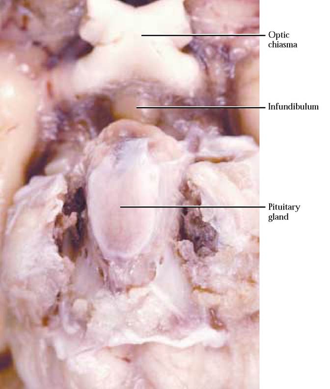
FIGURE 7-1 Hypophysis.
Figure 7-2 and Figure 1-21]. It produces a myriad of hormones, many of which control secretions of other glands.
PINEAL GLAND
The pineal gland projects from the dorsal surface of the brain [Figure 7-2 and Figure 7-3]. The hormone produced by this gland may play a role in the commencement of puberty.
THYROID GLAND
The thyroid gland is paired and lies on either side of the trachea just posterior to the larynx [Figure 7-4]. Commonly, the lobes of this gland are connected by a delicate, narrow band of tissue called the isthmus, difficult to find. Hormones produced by this endocrine tissue are involved in a wide range of metabolic activities, tissue maturation, sexual maturation, and other essential cellular activities such as energy utilization.
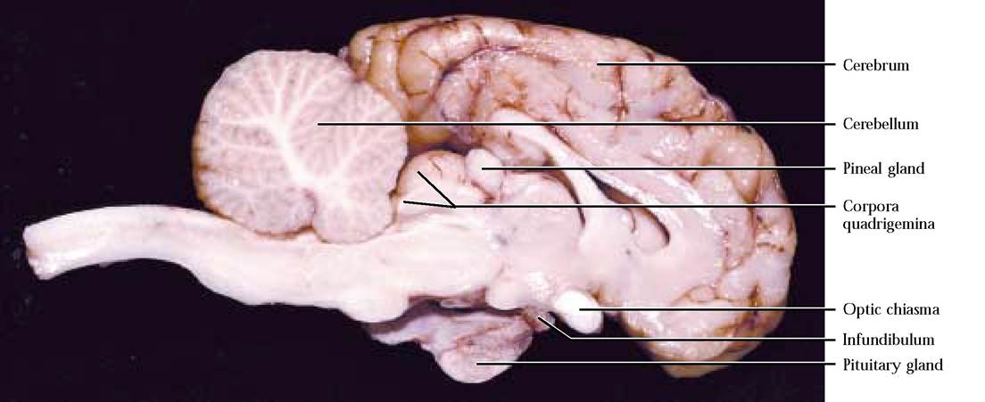
FIGURE 7-2 Brain: sagittal section.
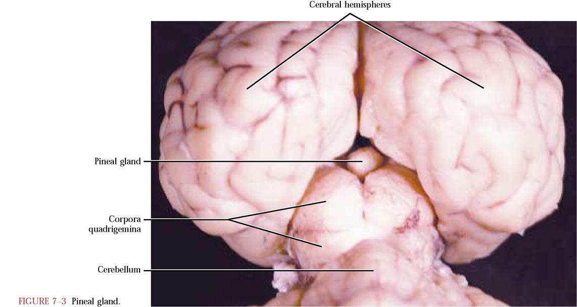
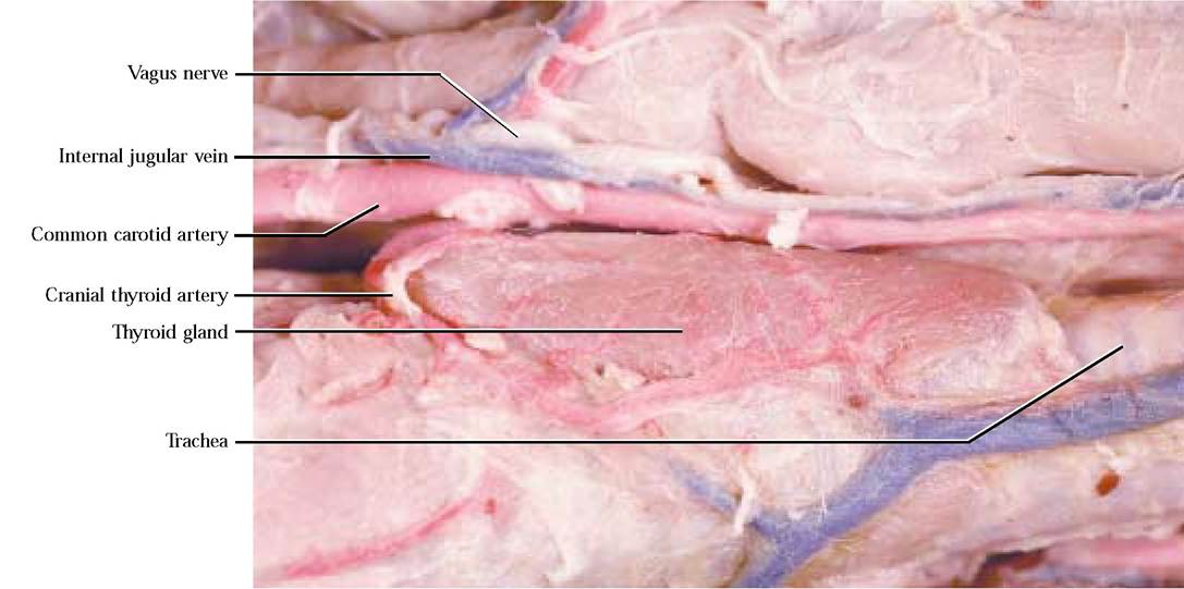
FIGURE 7-4 Thyroid gland.
PARATHYROID GLAND
THYMUS GLAND
The four very small parathyroid glands are embedded in the dorsal surfaces of the thyroid glands but are not always easily identifiable [Figure 7-5]. Their hormone secretion is critical to calcium balance.
The thymus is a lobular gland that is very obvious in young mammals but may be difficult to find in older cats [Figure 7—6]. It lies on the ventral aspect of the trachea and may extend over the surface of the heart. During the juvenile
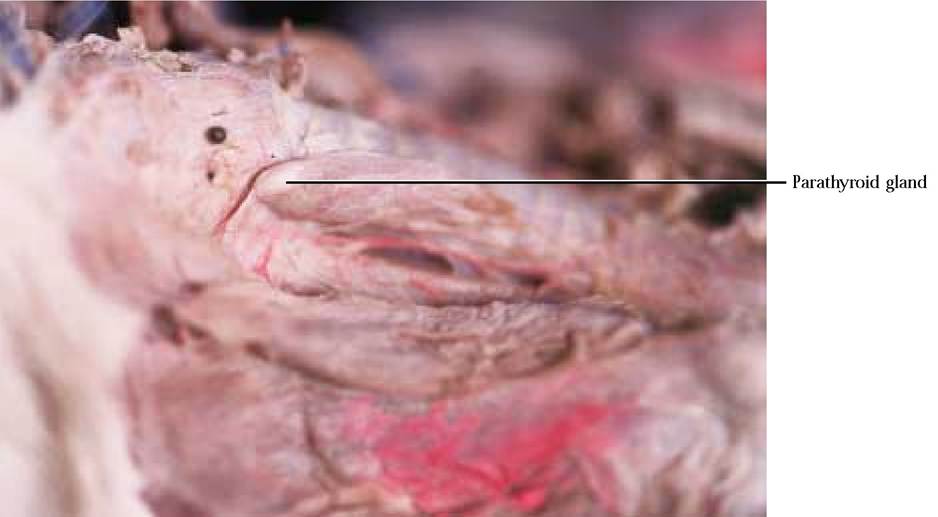
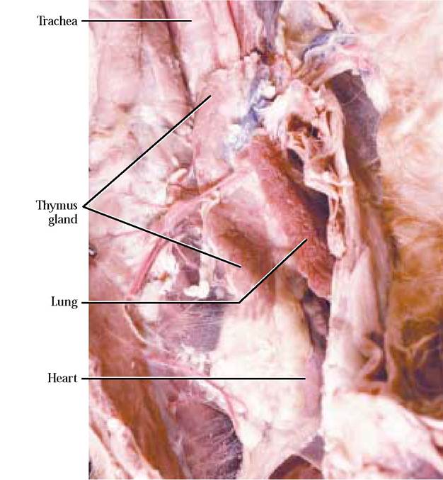
FIGURE 7-6 Thymus gland.
period of mammalian life, the thymus functions as a source of specialized leukocytes that migrate to other tissues to participate in immune activities.
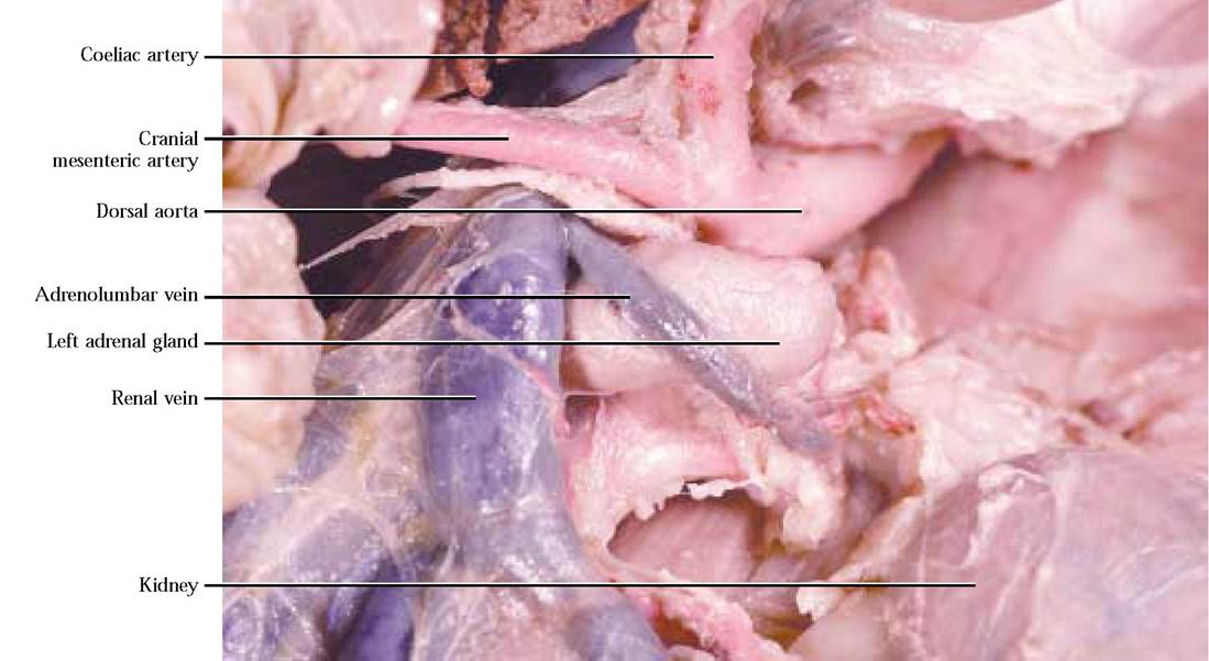
ADRENAL GLANDS
The small ovoid adrenal glands are located a short distance anteriomedial to the kidneys [Figure 7—7]. In contrast, the adrenal glands in the human adhere to the cranial surface of the kidneys. This organ is constructed of two areas, the outer cortex and the inner medulla. The hormones secreted by the cortex regulate a number of metabolic activities while those of the medulla intensify and prolong the characteristic syndrome stimulated by the sympathetic portion of the autonomic nervous system.
PANCREAS
The pancreas is a lobular gland located in the mesoduodenum and part of the gastrosplenic portion of the greater omentum [Figure 7-8]. This gland consists of two types of tissue, one that functions as endocrine tissue and the other that secretes chemicals involved in digestion. Hormones produced by the endocrine portion affect carbohydrate metabolism.
OVARIES
TESTES
The small, oval ovaries of the female reproductive system are located within the peritoneal cavity and suspended from the dorsal body wall [Figure 7—9].
The endocrine portion of the ovary produces female sex hormones that affect sexual and reproductive behavior.The testes, the male reproductive organs, located in the scrotum produce sex hormones affecting sexual and reproductive behavior [Figure 7—10].
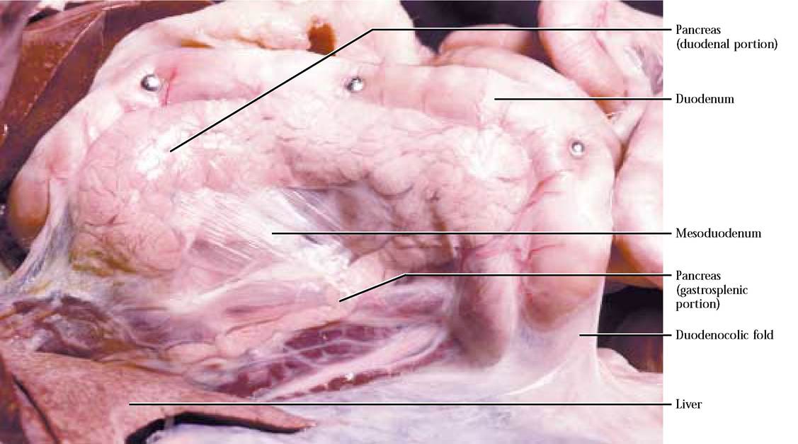
FIGURE 7-8 Pancreas.
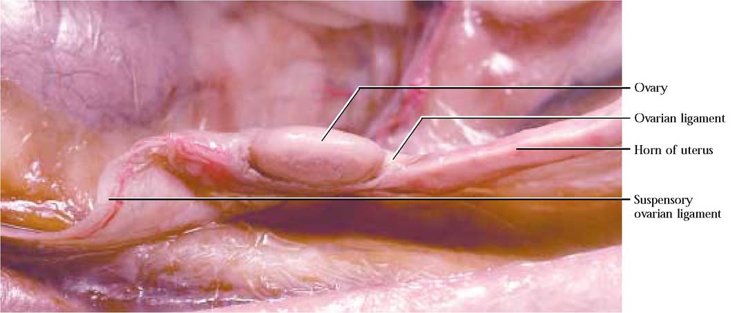
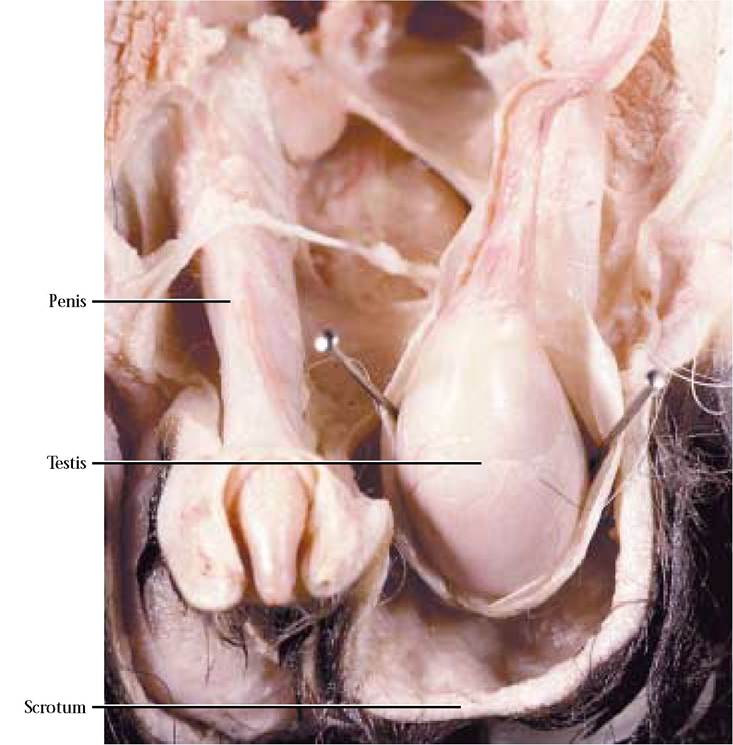
FIGURE 7-10 Testis.
OTHER ENDOCRINE TISSUES
Tissues in the kidney and digestive organs produce hormones whose activities tend to be more localized and confined to the functions of the organs in which they are produced.