Urogenital System
As the name of this system suggests, it is a combination of two different systems. In mammals, as in many other vertebrates, the excretory and reproductive systems are closely interrelated, not only anatomically, Eutalsoembryologically.
Inparticulapmostoftheterminalductworkinmalesiscommontobothsystems. Development of the reproductive systems in both sexes begins with an indifferent stage. Genetically, the sex is determined, but in the early stages of development there are no morphological differences in the gonads and external genitalia. Under the influence of testosterone, normal development of the reproductive system of male mammals progresses, whereas female reproductive structures develop in the absence of testosterone.In spite of the interrelationships of the excretory and reproductive systems, the functions are worlds apart. In both sexes, the excretory system functions in maintaining the homeostatic balance of fluids, electrolytes, glucose, hormones, proteins, metabolic waste products, and other chemical substances. This balance is mediated through filtration, reabsorption, secretion, and elimination of excess chemicals above normal blood threshold levels. The structure and function of the excretory system in the two sexes is virtually identical from the kidneys to the urethra. In males, the pre-prostatic urethra is purely urinary while the rest of this tube functions as a part of both the excretory and reproductive systems. In females the entire urethra is urinary.
The functions of the testes and the accessory glands of the male reproductive system include sex hormone and semen production (sperm and associated glandular fluids). Hormones are important to maintain “maleness” and continued stimulation of semen production. While urine passes from the urinary bladder to the outside by way of the entire length of the urethra, semen, on the other hand, travels only along that part of the urethra distal to the prostate gland to the outside.
Deposition of the seminal fluid into the female reproductive tract is one of the functions of the penis.The functions of the ovaries are production of sex hormones and oocytes and secretions by accessory glands in the female reproductive system. The system of “tubes” in the female reproductive apparatus is adapted for the receipt of semen from the male during intercourse, movement of the sperm to effect fertilization, and subsequent possible implantation and continued nourishment and maintenance of the developing embryo in the uterus.
EXCRETORY SYSTEM
Kidney
The kidney is the fundamental organ of this system. The primary functional units of this organ are the nephrons. The nephron consists of a cup-like Bowman's capsule with an elongate tubule extending from it. A specialized arterial capillary, the glomerulus, is intimately associated with the capsule. Also associated with the tubules of the nephron are other capillaries, numerous blood vessels, and nerves. The volume of blood that courses through the
kidneys of mammals is impressive, e.g., in humans, the total blood volume of about 5 liters filters through the glomerulus approximately every 40 minutes. The end product of the filtration, reabsorption and secretion of the nephron is urine. In humans, in spite of the large volume of filtrate, 180 liters, resulting during a 24 hour period, only about 1 liter of urine is eliminated.
The kidneys are paired, retroperitoneal (“behind” the parietal peritoneum), organs that are surrounded by fat deposits in the dorsal portion of the lumbar region. Notice that the position of the right kidney is slightly more caudal than the left due to the posterior extension of the caudate lobe of the liver on the right side. Take note of the hepatorenal ligament extending between the caudate lobe and the right kidney. It has no counterpart on the left side, therefore be careful to conserve this membranous structure [Figure 3-8 and Figure 6—1].
In order to observe the gross internal anatomy of the kidney, make a slit through the parietal peritoneum on the left side.
Carefully separate the left kidney from the surrounding fat, taking care to expose it sufficiently to allow you to make a mid-frontal cut through the kidney. In life the kidney resembles a large kidney bean in color and in shape. The medial indentation is the hilus [Figure 6—1]. Through this region passes the expanded proximal end of the ureter, the renal pelvis, renal arteries and veins, and nerves. Notice the tough, whitish, fibrous connective tissue encapsulating the kidney, the renal capsule. To better view this capsule, carefully peel it back, without removing it [Figure 6—2]. The outer narrow band of lighter tissue in the section is the granular cortex. The central darker region is the striated medulla. The glomerulus and portions of the nephron tubule are found in the cortex while other tubular regions of the nephron as well as collecting ducts are found in the medulla.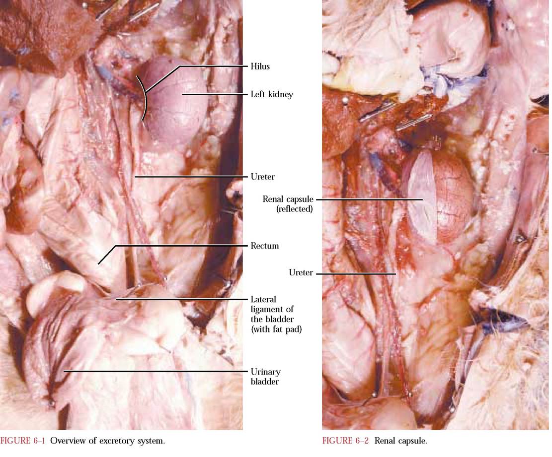
The medulla of the kidney in the cat consists of a single pyramid with its base abutting the cortex and its vertex opening as the papilla into the renal pelvis, the expanded proximal end of the ureter. The renal pelvis is located within the renal sinus, the space surrounding the renal pelvis [Figure 6-3]. Fat and blood vessels may be seen in the renal sinus. The structure of the human kidney is quite similar. The major difference is that the human kidney is multipyramidal with each of the pyramids terminating in a renal papilla that projects into areas known as calyces. The calyces, in turn, open into the renal pelvis.
Ureters, Urinary Bladder, and Urethra
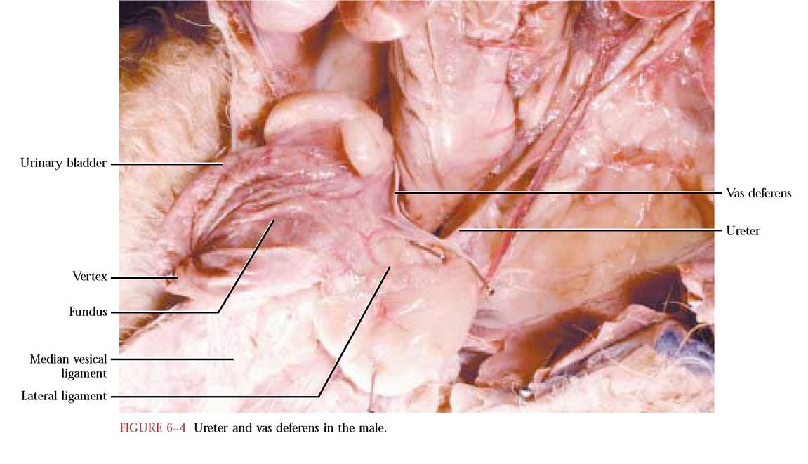
Ureters are tubes with muscular walls leading from the kidney to the urinary bladder. The ureter begins as the renal pelvis located within the sinus of the kidney, courses caudally in a retroperitoneal position, and passes through the lateral ligament of the urinary bladder to enter the urinary bladder [Figure 6-1]. The ureteral openings along with the urethral opening delineate a triangular area in the base of the bladder.
Carefully pick away the connective tissue and fat covering the left ureter exposing it from the hilus to the urinary bladder. Do not destroy the entire left lateral ligament of the bladder. In males, the vas deferens coils around the ureter at the base of the urinary bladder. Do not damage the vas deferens [Figure 6-4].The saclike organ, the urinary bladder, is a reservoir for urine. The walls of the bladder from the luminal side to the outer surface consist of several layers- a mucosa, a submucosa, a muscular layer and the serosal layer. When it is empty or in the relaxed state, it will appear small and have a muscular tone. As urine enters via the ureteral openings, the bladder expands to accommodate the incoming fluid. It may become greatly extended with urine and appears more saclike. When the bladder is relaxed, its inner walls are folded into rugae. When filled, the rugae disappear because of stretching caused by accumulating urine. The free domed end of the urinary bladder is the vertex while the attached caudal end is the fundus [Figure 6-4]. Remember that the bladder is held in position ventrally by the median ligament and on each side by the lateral ligaments, containing pads of fat.
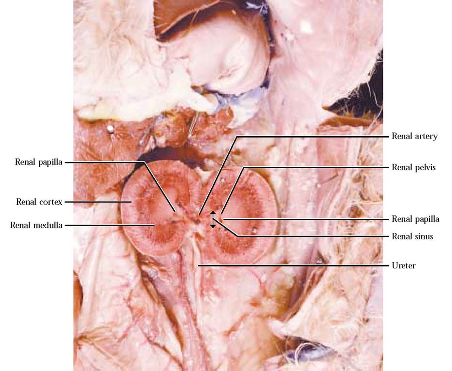
FIGURE 6-3 Internal anatomy of the kidney.
Urine leaves the urinary bladder by way of the tubular urethra [Figure 6-7 and Figure 6-15]. Like the ureters and the urinary bladder, the walls of the urethra consist of similar tissue layers. Since this tube lies primarily in the pelvic canal, it will be seen during the dissection of the reproductive system.
In humans, the urinary system, with the exception of the aforementioned kidney differences, is very similar.
FEMALE REPRODUCTIVE SYSTEM
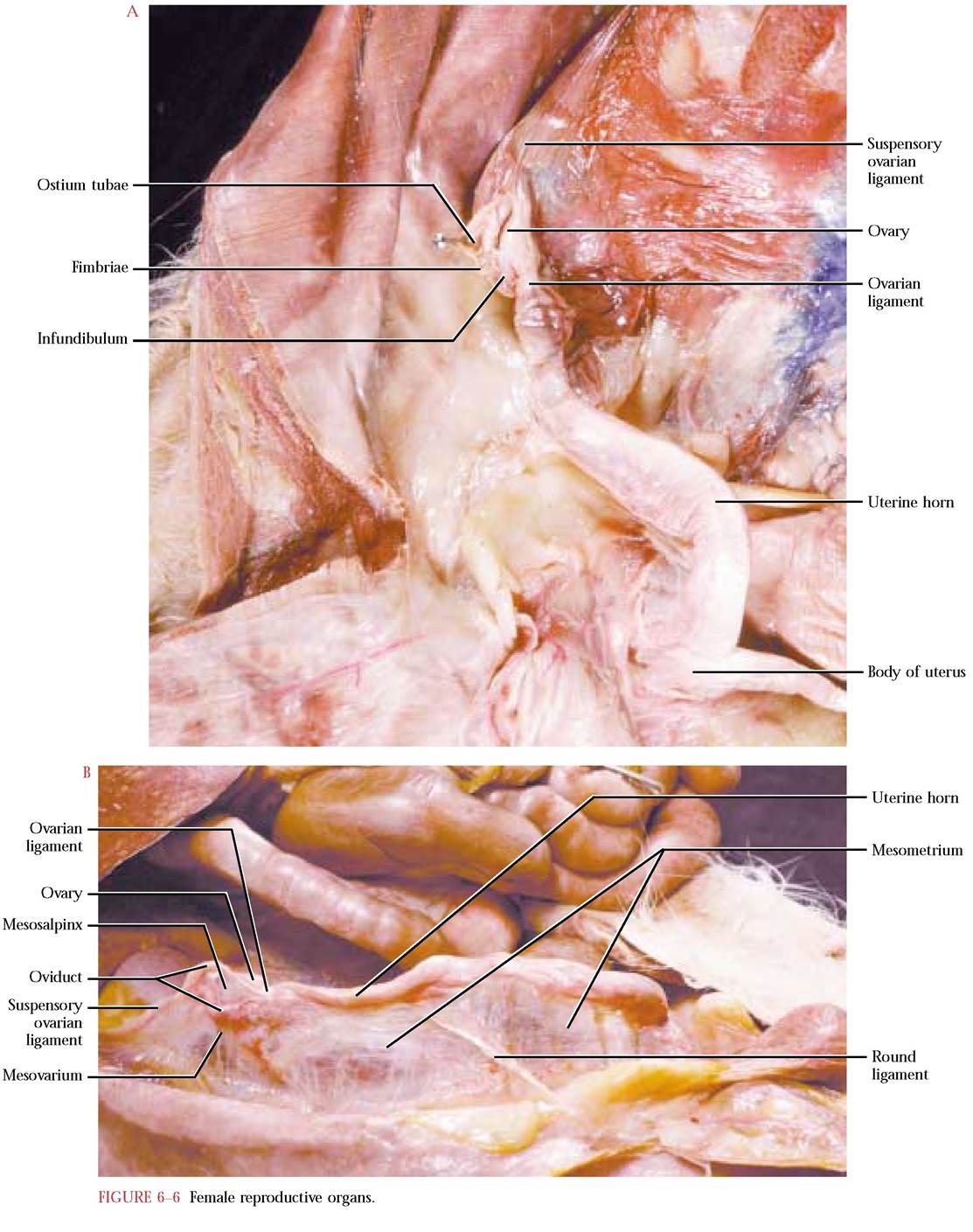
The primary reproductive organs are the ovaries. Ovaries are small, oval organs, suspended from the dorsal body wall cranially by the suspensory ovarian ligament and caudally by the ovarian ligament connecting it to the cranial end of the uterine horns.
Remember that the ovaries are further supported by a portion of the broad ligament, the mesovarium [Figure 6-5, Figure 6-6A, and Figure 6-6B].In reality, the ovaries are not physically connected to the tubes associated with the female reproductive system. Therefore, when the oocytes rupture through the serosa of the ovary they are swept into the oviducts or uterine tubes. In mammals, fertilization occurs in the upper third of the oviducts. The small, coiled oviducts lie lateral to the ovary and are suspended by another portion of the broad ligament, the mesosalpinx. The expanded proximal end of the oviduct, the infundibulum, is hood-like and wraps around the ovary laterally and opens medially by way of the ostium tubae. Along its edges are small, finger-like projections, the fimbriae, whose movements are responsible for the sweeping of the oocyte into the oviduct. To observe these structures, carefully reflect the ovary and uterine tubes laterally [Figure 6-6A and Figure 6-6B]. To appreciate the ostium tubae, carefully separate the edges of the infundibulum and insert a probe between them.
The uterine tubes merge into the larger diameter horns of the uterus (uterine horns). The two uterine horns fuse to form the body of the uterus and give the impression of a Y-shaped organ [Figure 6-5 and Figure 6-6A]. Successful fertilization results in implantation of early embryos in the uterine horns where development continues for nine to ten weeks, leading to birth of kittens, small versions of the adult. Recall that the mesometrium of the broad ligament supports the horns of the uterus. Note the round ligament present in the mesometrium. It is homologous with the gubernaculum in the male (see page 119).
To view the distal portion of the reproductive system, it is necessary to expose the pelvic cavity. While being cautious to avoid the median ligament of the bladder and blood vessels on the undersurface of the abdominal window, carefully make about a one-inch centrally located horizontal cut along the pelvic rim.
Insert your index finger and palpate with your thumb externally to ascertain the position of the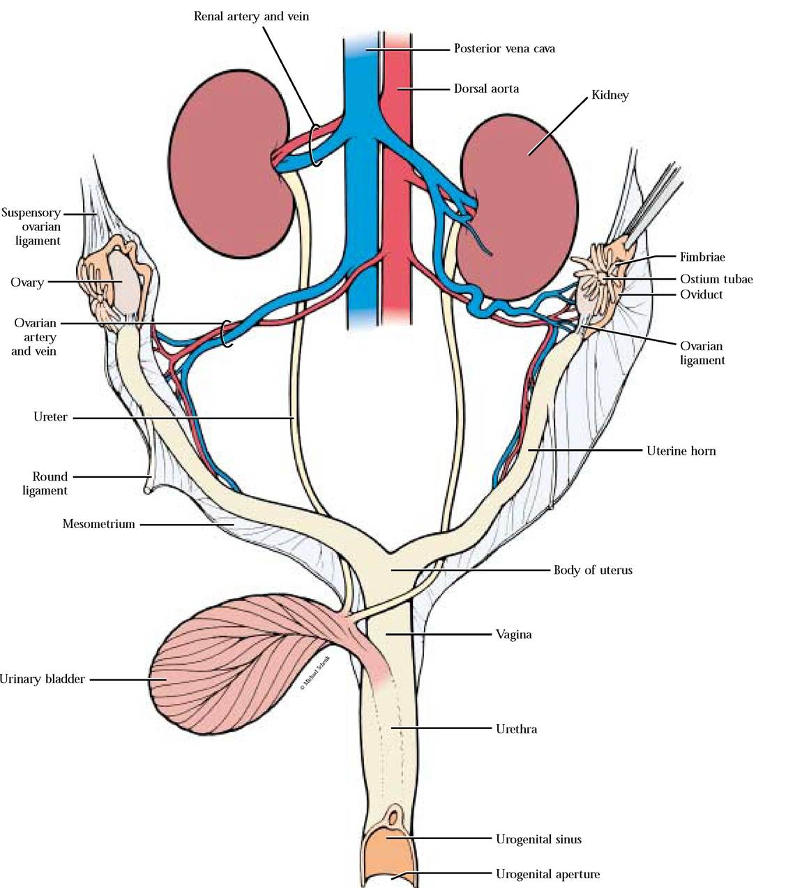
pubic symphysis that will feel like a shallow groove. This should correspond with the juncture of the gracilis muscles of the hindlegs which appears as a raphe (line) between the muscles overlying the symphysis. With a sharp scalpel make an incision through the line forming the muscle juncture. Often, the incision will follow the margin between the paired pubis and ischium closely and results in a clean separation of these bones. If that is not the case, with a pair of bone cutters, cut through the symphysis of the os coxae. Now, grasp the legs and break through the symphysis, exposing the organs of the pelvic cavity. Carefully clean the exposed area of fat and connective tissue, being especially careful not to remove associated blood vessels and nerves [Figure 6-7].
To observe the anatomy of the distal portion of the urogenital system, with a pair of scissors beginning at the urogenital opening, make a cut through the lateral wall of the canal. While making this short cut, continue until you observe the opening of the urethra and vagina into the urogenital sinus. Do not cut further [Figure 6-8].
The body of the uterus tapers distally to form the cervix, the neck-like region of the uterus, which protrudes into the vagina [Figure 6-7]. The cervix can be palpated externally as a sphincter-like region. The vagina extends from the cervix to the urogenital sinus where it opens as the vaginal orifice along with the urethral orifice, the opening of the urethra, thus serving as a common canal for the genital and urinary systems.
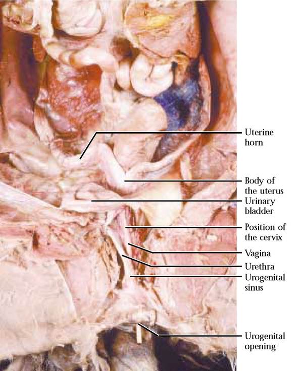
FIGURE 6-7 Distal female urogenital system.
To observe these openings and other features of the urogenital system, reflect the cut ventral half of the canal. The urogenital sinus is quite long in the cat and opens to the outside through the urogenital aperture. In the cat, the labia are slight skin folds situated laterally around the urogenital aperture, that are not easily identifiable. Notice the small papillate clitoris resting in a shallow, midventral depression [Figure 6-9]. This organ is partially homologous with the penis.
While describing the anatomy of the digestive system, the terminal portion of the rectum was not observable until the pelvic canal was exposed [Figure 6-9]. Notice its position dorsal to the uterus as it continues to the outside through the anus. The anal glands are located on either side of the anus and open into the rectum. Cats use the secretions of these glands to mark their territories and also
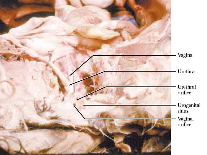
FIGURE 6-8 Female urogenital sinus with associated openings.
advertise their sex and sexual condition.
If a female cat is either pregnant, just given birth or suckling young, the paired mammary glands with their nipples extending along either side of the midventral line, will be very prominent [Figure 6-10 and Figure 6-11].
The anatomy of the human female is very similar with some minor differences. Among these are the absence of uterine horns, two pairs of labia, majora and minora, a more prominent clitoris, and a cleftlike area between the labia minora, the vestibule of the vagina, where the urethra and the vagina open to the outside. Note that this condition is different from the female cat where there is a well defined urogenital sinus. Some further specialization of supporting ligaments is evident.
MALE REPRODUCTIVE SYSTEM
The primary reproductive organs in the male are the testes, egg shaped organs that are suspended externally within the scrotum, an obvious hairy sac protruding posteriorly just ventral to the anus. Take note also of the position of the sheath of the penis with its opening ventral to the scrotum. One of the major problems encountered with the male reproductive system is knowing where the spermatic cords leave the abdominal cavity through the abdominal wall and realizing that they lie in the fat of the inguinal (groin) area and can be cut or destroyed in the wink of an eye. This is the reason that you were warned
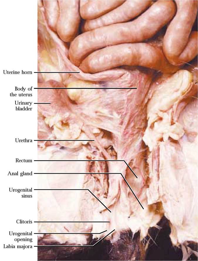
FIGURE 6-9 Terminal female urogenital system.
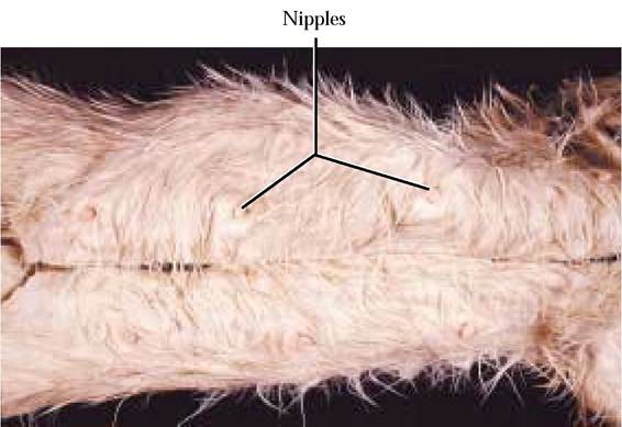
FIGURE 6-10 External features associated with mammary glands.
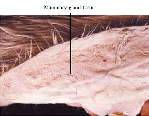
FIGURE 6-11 Internal mammary glands.
to avoid removing fatty tissue in this region during the skinning and initial connective tissue cleaning process [Figure 6-12].
The dissection of the male reproductive system is somewhat more difficult and time consuming than the female. Begin by cautiously making a small cut (1/8 inch) through the skin of the posterior wall of the scrotum. Carefully work the scissors under the skin and continue the incision to the anterior limit of the testis. Repeat the process on the other side. Now, carefully peel the scrotum laterally to expose the testis on both sides. Notice the abundant connective tissue, the cremasteric fascia, stretching between the inner wall of the scrotum and the white sac, the cremasteric pouch, surrounding the testis [Figure 6-13]. You will observe that the pouch abruptly narrows into a tubelike structure, the spermatic cord, at its anterior end. Within the spermatic cord lies the vas deferens, the spermatic artery and vein, lymphatic vessels, and nerves.
Leave one of the testes held in place by cremasteric fascia within the scrotum, but gently remove the other by loosening the cremasteric fascia. Do not cut the spermatic cord and detach the testis from the body of the cat; leave it attached. The organ you now observe is the testis enclosed within the cremasteric pouch. The pouch and its contents have the same relationship as any of the major ventral body cavities to the body proper, e.g., the abdominopelvic, pericardial or pleural cavities (see Figure 3-3A and Figure 3-3C). The tough, outer layer of the cremasteric pouch, the tunica vaginalis communis, is analogous to the body proper. Carefully snip through this outer covering and then continue the cut to expose the structures suspended within the sac. Notice the space between these enclosed structures and the sac. It is known as the vaginal cavity and is analogous to the potential space of a ventral body cavity. The inner lining of the pouch is the tunica vaginalis propria—parietal layer and is spatially analogous to the parietal layer of a ventral body cavity. This layer comes together to form the mesentery-like mesorchium and then spreads over the surface of the structures, suspended within the vaginal cavity, as the tunica vaginalis propria—visceral layer, spatially analogous to the visceral layer of a ventral body cavity [Figure 6-14]. The mesorchium can be demonstrated by gently grasping the structures of the spermatic cord and pulling them medially [Figure 6-15]. This thin, mesentery-like membrane anchors the vas deferens, blood and lymph vessels and nerves in place within the cord. The two layers of the tunica vaginalis propria can be seen as the inner shiny layer of the cremasteric pouch (parietal layer) and the outer shiny layer covering the testis (visceral layer).
A band of highly convoluted tubules, the epididymis, adheres to the dorsal portion of the testis. Observe the free anterior end, the head, the middle portion, the body, and the posterior tail of the epididymis. The vas deferens begins its journey as a somewhat convoluted tube from the tail of the epididymis and enters the spermatic cord, held in place by the mesorchium [Figure 6-15].
The descent of the testes in most male mammals has a very interesting developmental history. The testes begin their life in the abdominal cavity, but attached to the inner portion of the scrotum by a long band of connective tissue called the gubernaculum that passes through the lower abdominal wall on its way to the scrotum. As the fetus matures, the gubernaculum contracts, pulling the testes into the scrotum. As the testes move into the scrotum they are accompanied in their journey by a small bit of the peritoneal cavity (vaginal cavity) along with the parietal and visceral peritoneal layers. So, really, it is not surprising that the organs suspended within the vaginal cavity have very similar relationships to the space and membranes of the peritoneal cavity. To see the gubernaculum, while holding the testis, gently pull the cremasteric pouch wall at the posterior end of the testis and observe the short, tough fibrous band holding the tail of the epididymis tightly against the inner wall of the pouch [Figure 6-12 and Figure 6-15].
Now, very carefully separate the spermatic cord from surrounding tissues and trace it anteriorly to an opening piercing the abdominal wall, the external inguinal ring. The cord passes through a short inguinal canal and the vas deferens enters the abdominal cavity through the internal inguinal ring. Notice also that the spermatic artery and vein enter and exit the spermatic cord at this point. Follow the vas deferens as it courses dorsally around each of the ureters and urinary bladder [Figure 6-4 and Figure 6-15].
The next dissection should be completed preliminary to further work. Grasp the opening of the sheath of the penis with a pair of forceps and make a cut through the anterior wall of the sheath with scissors [Figure 6-15]. Continue the cut through the connective tissue along the shaft of the penis to expose it. Take care to avoid cutting into the penis itself.
To facilitate observation of the urethra, glands and other associated parts of the system, it is necessary to cut through the symphyses of the os coxae. As in the female, carefully make about a one inch cut through the abdominal wall along the rim of the pelvis, avoiding the median ligament of the bladder and blood vessels associated with the inner portion of the abdominal window cut when the cat was opened. Carefully palpate the slight depression between the pubes and with a sharp scalpel, cautiously make an incision, beginning with the depression and following the raphe between the gracilis muscles. Remember that the cut through the raphe will be quite deep. Now grasp the hindlegs and reflect them laterally to complete the separation of the two os coxae. It may be necessary to complete the separation by carefully cutting through the ischiatic symphysis with the scalpel. At all times be careful not to cut into organs lying within the pelvic canal.
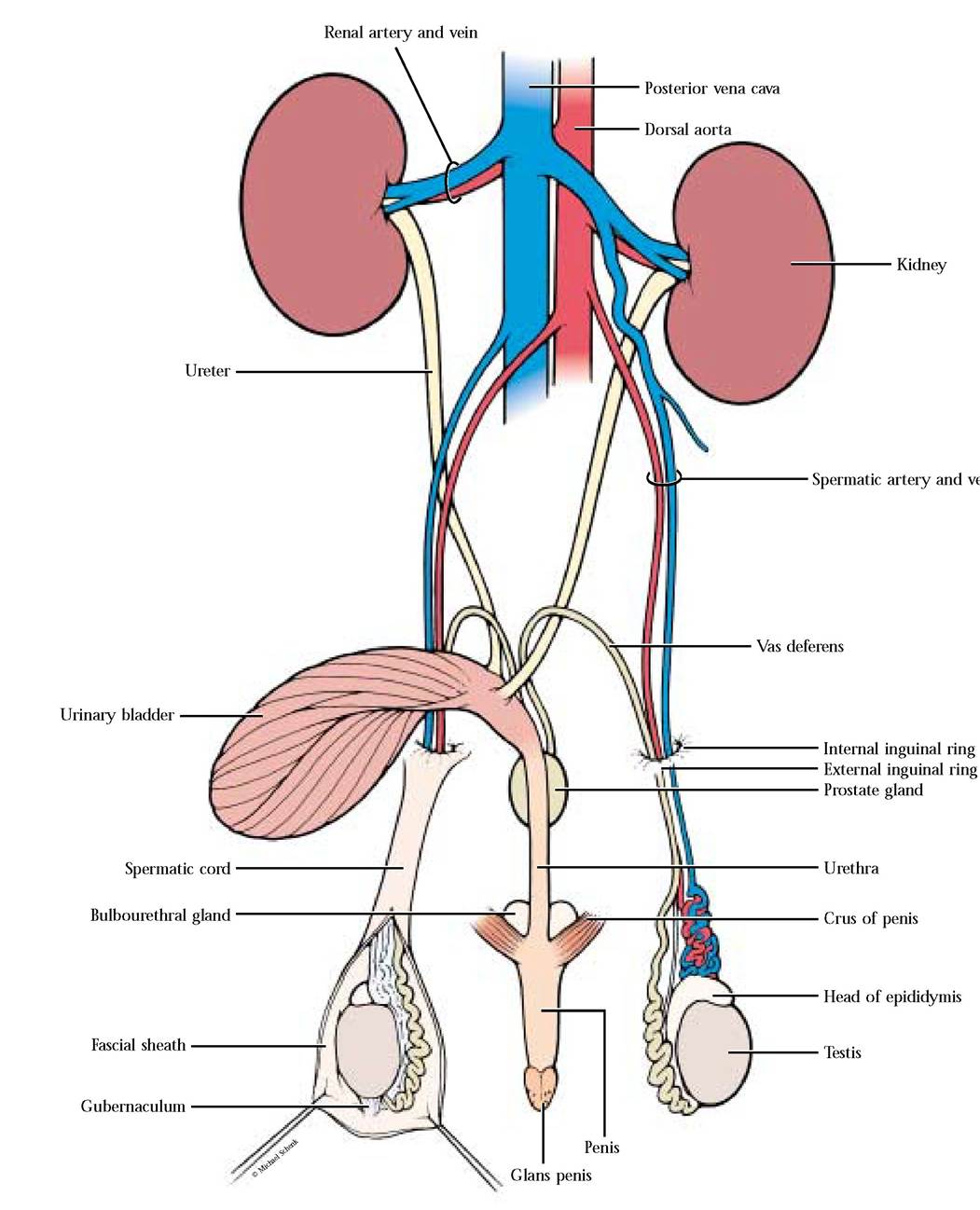
With some luck and good dissection you should be looking into the canal. Begin cleaning connective tissue from the tube leading from the urinary bladder, being careful to conserve associated blood vessels and nerves in the process. As you proceed distally an enlarged, whitish mass, the prostate gland, will be encountered. At this point the vasa deferentia connect to the tube. That part of the tube extending from the urinary bladder to the prostate is the urethra and carries only urine. That part of the tube distal to the prostate is the urogenital canal that continues to the tip of the penis where it opens to the outside through the urogenital aperture. Both urine and semen are carried in the urogenital canal [Figure 6—15].
The distal portion of the urogenital canal is surrounded by a column of erectile tissue, the corpus spongiosum, that lies along the caudal surface of the penis when not erect. The distal portion of this erectile tissue caps the tip of the penis as the conical glans penis. The urogenital aperture opens at the tip of the glans. Notice the pocket of skin, the prepuce, that encloses the glans [Figure 6—14].
A pair of erectile columns of tissue, the corpora cavernosa, lie along the cranial surface of the penis, when it is not erect. All three of these columns possess blood sinuses that bring about erection under the influence of sexual excitement. The proximal ends of the corpora cavernosa are attached to the ischia by bands of tough connective tissue called the crura of the penis (singular: crus). Each crus is covered ventrally by the ischiocavernosus muscle. Dorsal to each of the crura is a bulbourethral gland (Cowper's gland) that secretes lubricating fluid into the urogenital canal during sexual excitement [Figure 6-15].
As in the female, the male also possesses anal glands that are situated near the terminal end of the large intestine, the rectum, near the anus [Figure 6-15]. These serve a similar function in males. Perhaps you have seen large cats, e.g., lions and tigers, in those wildlife documentaries on television as they back up to a bush or shrub and spray fluid on them. The fluid is forcefully ejected from their anal glands. Unneutered male domestic cats do something similar on furniture and other objects in their home territories, by the way, leading to odoriferous surroundings.
The male reproductive system in human males is quite similar to the cat, again with some exceptions. The inguinal canal in the human is considerably longer than in the cat. Since the abdominal wall is thinner in this region, it may be the site of an abnormal condition known as an inguinal hernia. The distal ends of the vasa deferentia enlarge into ampullae where they are joined by the ducts of the seminal vesicles to form the ejaculatory ducts, that in turn connect to the urogenital canal. The seminal vesicles secrete part of the fluid volume of the semen. The penis in humans is not contained within a sheath but hangs freely, attached proximally to the pubic symphysis by way of the crura.
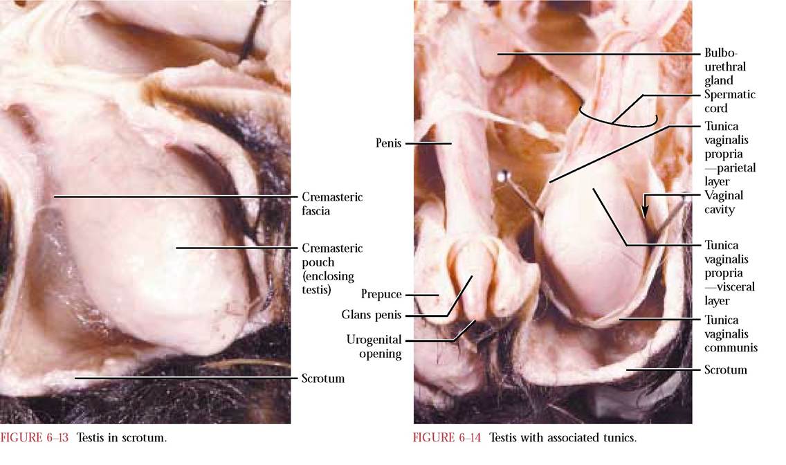
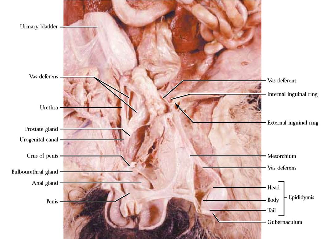
FIGURE 6-15 Male urogenital system.