ERYTHROCYTES
1. What chemical atom associated with hemoglobin binds loosely and reversibly with oxygen? How many molecules of O2 can be transported by one molecule of hemoglobin?
2.
What is the valence of iron before and after its binding with oxygen?3. What are methemoglobin, myoglobin, and carbon monoxyhemoglobin, and how do they differ from hemoglobin?
4. What is the average concentration of hemoglobin in the blood of domestic animals?
5. What is the physiologic name for the production of erythrocytes?
6. Where does RBC production occur during the postnatal, growth, and adult periods?
7. How does reticulocyte presence relate to lifespan of erythrocytes?
8. What substance controls the rate of erythropoiesis? Where is it produced?
9. How long does it take for new RBCs to enter the circulation after their formation begins?
0. If there are 7 million RBCs in each microliter of cow blood, how many would there be in 1 mL?
11. What are advantages of a discoid RBC shape? What is tolerance to RBC shape change known as?
2. Which domestic animal has the largest RBC? The smallest?
3. Which one of the erythrocyte indices is related to an erythrocyte’s volume? What is the unit of expression? What is the unit of expression for the amount of hemoglobin in each RBC?
Hemoglobin and Its Forms
The principal component of erythrocytes is hemoglobin (Hb), which makes up about one-third of the erythrocyte content, the remainder being water and stroma (structural components). The hemoglobin molecule (Figure 3-7) has a molecular weight of about 67,000 and is composed of four heme groups combined with one molecule of globin (the protein component). Globin is composed of four polypeptide chains, each containing one of the heme groups. Each heme group contains an iron atom that combines loosely and reversibly with one oxygen molecule.
Therefore, one molecule of hemoglobin contains four molecules of oxygen. The iron atom of heme has a valence of +2 (Fe2+, ferrous) regardless of whether molecular oxygen is combined with it. Because of the presence of hemoglobin, blood can transport about 60 times more oxygen than would be possible by its simple solution. Certain conditions cause the ferrous iron of heme to be oxidized to its ferric state. In one such condition, nitrate poisoning, the hemoglobin formed is known as methemoglobin and it cannot transport oxygen. Another abnormal form of hemoglobin is carbon monoxyhemoglobin (sometimes called carboxyhemoglobin). As the name implies, carbon monoxide occupies the site normally occupied by oxygen. Hemoglobin has an affinity for carbon monoxide that is about 200 times greater than its affinity for oxygen. Thus, small concentrations of carbon monoxide compete more favorably for sites on Hb than normal concentrations of oxygen.
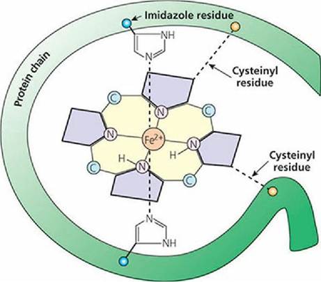
■ FIGURE 3-7 Schematic representation of one heme group and its associated polypeptide chain. Four of these combinations, at different orientations to each other, make up hemoglobin. The heme is held to its specific polypeptide chain (one of four in the protein globin) by cysteine (an amino acid) bridges and by bonding of the iron to imidazole groups of histidine (an amino acid). Molecular oxygen binds with iron. (Modified from Conn EE, Stumpf PK. Outlines of Biochemistry. New York: John Wiley & Sons, 1963.)
The hemoglobin of muscle is known as myoglobin. It differs from hemoglobin in that it has only one polypeptide chain and one associated heme group, so it can only combine with one molecule of oxygen instead of four. The concentration of hemoglobin in the blood of domestic animals averages about 12 g/dL (Table 3-2).
| TABLE 3-2 AVERAGE VALUES FOR SEVERAL BLOOD VARIABLES" | ||||||
| ANIMAL | ||||||
| VARIABLE | HORSEb | COW | SHEEP | PIG | DOG | CHICKEN |
| Total RBC∕μL blood (?106) | 9.0 | 7.0 | 12.0 | 6.5 | 6.8 | 3.0 |
| Diameter of RBC (μm) | 5.5 | 5.9 | 4.8 | 6.0 | 7.0 | elliptic.7 ? 12 |
| PCV (%) | 41.0 | 35.0 | 35.0 | 42.0 | 45.0 | 30.0 |
| Sedimentation rate.(mm∕min) | 2-12∕10 | 0∕60 | 0∕60 | 1- 14∕60 | 6- 10∕60 | 1.5-4∕60 |
| Hemoglobin (g∕ dL) | 14.4 | 11.0 | 11.5 | 13.0 | 15.0 | 9.0 |
| Coagulation time (capillary tube method; min) | 2-5 | 2-5 | 2-5 | 2-5 | 2-5 | C |
| Specific gravity (g∕ dL) | 1.060 | 1.043 | 1.042 | 1.060 | 1.059 | 1.050 |
| Plasma protein (g∕ dL) | 6-8 | 7 8.5 | 6-8 | 6.5 8.5 | 6-7.8 | 4.5 |
| Blood pH (arterial) | 7.40 | 7.38 | 7.48 | 7.4 | 7.36 | 7.48 |
| Blood volume (percent of body weight) | 8-10 | 5-6 | 5-6 | 5-7 | 8-10 | 7-9 |
| Mean corpuscular volume (MCV; fL) | 45.5 | 52.0 | 34.0 | 63.0 | 70.0 | 115.0 |
| Mean corpuscular hemoglobin (MCH; pg) | 15.9 | 14.0 | 10.0 | 19.0 | 22.8 | 41.0 |
| Mean corpuscular hemoglobin concentration (MCHC; %) | 35.0 | 33.0 | 32.5 | 32.0 | 34.0 | 29.0 |
aData compiled from Swenson MJ.
Physiological properties and cellular and chemical constituents of blood. In: Swenson MJ, Reece WO, eds. Duke’s Physiology of Domestic Animals, 11th Ed. Ithaca, NY: Cornell University Press, 1993, and Jain NC. Essentials of Veterinary Hematology. Philadelphia, PA: Lea & Febiger, 1993.⅛ot blooded.
cSee Species Differences in Tests for Blood Coagulation.
Erythropoiesis
The production of erythrocytes is known as erythropoiesis. Before birth, erythrocyte formation occurs in the liver, spleen, and bone marrow. During the postnatal, growth, and adult periods, erythropoiesis is restricted almost exclusively to the bone marrow. It seems that most bones are involved in erythropoiesis and the axial and appendicular skeletons account for about 35% and 65% of RBC production, respectively. This pattern has been observed in 1-year-old beagle dogs and can vary in other animals. The axial skeleton includes almost all bones except those of the limbs, which belong to the appendicular skeleton. The erythrocytes are continually formed and destroyed. Considering the large number of RBCs in the blood, one should appreciate the dynamic aspect of this phenomenon. For example, approximately 35,000,000 erythrocytes are formed and destroyed in a 450-kg horse each second.
Erythrocytes are formed in the bone marrow from a foundation cell known as a rubriblast (see Figure 3-3). Several intermediate forms are recognized in the genesis of the erythrocyte (Figure 38). The distribution of these forms can be studied by preparation and examination of bone marrow smears. Just before the developing erythrocyte’s entrance into the circulation, the nucleus is expelled. The polyribosomes and ribosomes are retained and might still be apparent on stained smears for a day or so after their arrival in the circulation. If they are present, they are identified as reticulocytes because of the net-like appearance of the polyribosomes and ribosomes. Polyribosomes (polysomes) consist of several ribosomes joined together by the same messenger RNA molecule.
During periods of rapid RBC production, reticulocyte numbers can increase. Reticulocytes are usually present in the blood of animals when the lifespan of erythrocytes is less than,100 days. The dog is an exception. Adult ruminants, and especially horses, with longer RBC lifespans do not have reticulocytes in the circulating blood when in health. The nuclei of avian erythrocytes are not expelled before entry into the circulation and they persist throughout the life of the erythrocytes.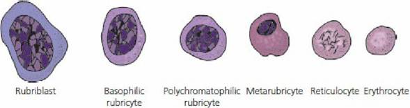
■ FIGURE 3-8 The stages of erythrocyte development.
The rate of erythropoiesis seems to be controlled by the tissue need for oxygen. Reduced oxygen at the tissue level results in the secretion of a hormone known as erythropoietin. Erythropoietin stimulates the bone marrow to begin formation of new erythrocytes. The lifespan of erythropoietin is less than 1 day; this short lifespan helps provide greater flexibility in the adjustment of erythrocyte numbers in order to regulate the tissue need for oxygen more precisely. New erythrocytes do not appear in the circulation until about 5 days after their formation begins. Thus, additional erythropoietin can be formed to allow continued production during the interim. When the new erythrocytes appear in the circulation, the tissue need for oxygen begins to be met and erythropoietin is no longer secreted.
Numbers
The number of erythrocytes can be determined by making known dilutions and counting the number of RBCs in a known volume using the counting chamber of a hemacytometer with the aid of a microscope. The Unopette© microcollection system (Becton Dickinson and Company, Franklin Lakes, NJ) is widely used for this purpose. In addition to erythrocytes, leukocytes and platelets can also be enumerated with this system. Using various multiplication factors (which make allowance for dilution and for the limited volume that is counted), the number of RBCs per microliter of blood can be determined.
More accurate determinations can be made using electronic counting equipment. A number of systems are available that are capable of counting erythrocytes, leukocytes, and platelets and of determining hemoglobin concentration. The cells are counted as they stream past a photoelectric cell in single file. A computer within provides printouts of means, ranges, and callouts for highs and lows. The erythrocyte indices are also calculated. Generally, there are about 7,000,000 RBCs per microliter of blood in the cow, pig, and dog (see Figure 3-2). More RBCs are seen for hot-blooded horses (9,000,000/pL) and for sheep (11,000,000/pL). Values for the goat are not given in Figure 3-2, but they average about 13,000,000/pL. See Appendix A for erythrocyte profiles (range and average) for each of the domestic animal species.Shape
Erythrocytes are generally considered to be discocytes, with some degree of concavity. The dog’s RBCs are typical biconcave disks, whereas the goat’s RBCs are more spherical. The camel has elliptical RBCs and the deer has RBCs that are somewhat sickle-shaped. The advantages of a discoid shape are (1) the provision of a larger surface area-to-volume ratio, (2) minimal diffusion distance, and (3) greater osmotic swelling (water intake) possible without threatening the integrity of the membrane.
The characteristic shape of erythrocytes is maintained by the molecular constitution of hemoglobin and by certain contractile proteins of the cell membrane. An altered shape, because of a difference in hemoglobin constitution, can result in disease such as sickle cell anemia in humans. A genetically induced substitution of the amino acid valine for the usual glutamic acid in the amino acid sequence of hemoglobin causes RBCs to assume a sickle shape, rather than the usual biconcave disk shape, when hemoglobin is deoxygenated. The altered shape makes the cells more vulnerable to destruction and anemia results.
Erythrocytes are tolerant of shape changes as they circulate.
Many variations are noted as they pass through the small lumen (duct) of capillaries or rebound from a collision with a vessel bifurcation (branch). This property of tolerance for shape change is known as plasticity.Size
Among the domestic animals, dogs have erythrocytes with the largest diameter (7 μm) and sheep and goats have those with the smallest (4-4.5μm). It seems that this was an adaptive feature for sheep and goats, because RBCs of the smallest size are found in greater numbers. Because the sheep and goat were commonly found in regions of high altitude, with lower oxygen concentrations, the available hemoglobin was placed in a greater number of smaller packages so that a greater surface area would be available for diffusion.
Erythrocyte Indices
The erythrocyte indices are determinations that are calculated after the erythrocytes (RBCs) have been enumerated and Hct and Hb concentrations determined. There are three indices and each relates to a value for a single RBC. Accordingly, the units are small and are shown for each as follows:
• Mean corpuscular volume (MCV) in femtoliters (fL); femto is one-quadrillionth (10-15).
• Mean corpuscular hemoglobin (MCH) in picograms (pg); pico is one-trillionth (10-12).
• Mean corpuscular hemoglobin concentration (MCH) in g/dL (deciliter) or g percent.
Derivations of values are as follows (exponent manipulations completed but not included):
Mean corpuscular volume
MCV = (Hct/RBC) ? 10
Example: Hct = 42%; RBC = 7 million^L
MCV(42/7) ? 10 = 60 fL
Mean corpuscular hemoglobin
MCH = ([Hb]/RBC) ? 10
Example: [Hb] = 14 g/dL; RBC = 7 million^L
MCH= (14/7) ? 10 = 20 pg
Mean corpuscular hemoglobin concentration
MCHC = ([Hb]/ Hct) ? 100
Example: [Hb] = 14 g/dL; Hct = 42%
MCHC= (14/42) ? 100 = 33.3%
The values for these indices are shown for each species in Figure 3-2. The indices are valuable aids in the diagnosis of various anemias.
Lifespan
The lifespan of erythrocytes varies with species. Reported values for horses are 140-150 days. In adult ruminants (cattle, sheep, and goats) erythrocyte lifespan varies from 125 to 160 days, in pigs from 75 to 95 days, in dogs from 100 to 120 days, and from 70 to 80 days in cats. The lifespan of erythrocytes in chickens is 20-30 days.
■