LEUKOCYTES
1. How are leukocytes classified? Where are the various cells produced? What do segmented and band cells refer to?
2. Which one of the leukocytes seems to have the longest lifespan?
3.
How do the numbers of red blood cells (RBCs) and white blood cells (WBCs) compare?4. Which WBC predominates in the horse, dog, and cat? In the pig, cow, sheep, and goat?
5. Describe the movement of neutrophils from the circulation to sites of inflammation.
6. What is a principal function for each of the leukocytes?
7. Which WBC is classified as a mononuclear phagocytic system cell? Which mononuclear phagocytic system cell is in a fixed position in the liver?
8. Which one of the WBCs becomes more numerous in certain types of parasitisms?
9. Differentiate between the functions of lymphocyte T cells and B cells.
0. What are plasma cells and megakaryocytes?
11. Differentiate among leukopenia, leukocytosis, and leukemia.
2. What is meant by absolute numbers of leukocytes?
3. Define phagocytosis, pinocytosis, and endocytosis.
Classification and Appearance
Leukocytes, or white blood cells (WBCs), are classified as either granulocytes, containing granules in the cytoplasm, or as agranulocytes, containing few if any granules in the cytoplasm. There are three types of granulocytes, named according to which component of the hematoxylin and eosin (H&E) stain (hematoxylin, basic and colored blue; eosin, acidic and colored red) is taken up by their granules. Neutrophils are neither markedly acidophilic nor basophilic and incorporate both basic and acidic components into their granules. Basophils only accept the basic (hematoxylin) component and eosinophils only accept the acidic (eosin) component. There are two types of agranulocytes: monocytes and lymphocytes. Granulocytes and monocytes are produced in the bone marrow from myeloid stem cells known as myoblasts and monoblasts, respectively.
Lymphocytes originate from a lymphoid stem cell, known as a lymphoblast, in lymph tissue, such as lymph nodes, spleen, tonsils, and various lymphoid clusters in the intestine and elsewhere. The different types of leukocytes are shown in Figure 3-2 for humans; there are many similarities with those in animals.
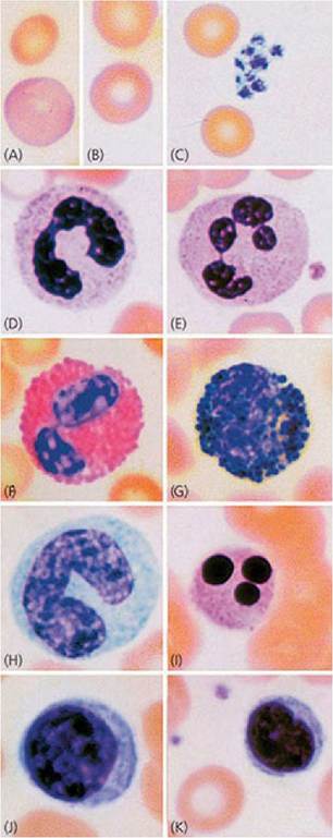
■ FIGURE 3-2 Cell types found in smears of normal peripheral blood: (A) polychromatophilic erythrocyte; (B) erythrocyte (mature); (C) platelets; (D) band neutrophil; (E) neutrophil (mature); (F) eosinophil; (G) basophil; (H) monocyte; (I) degenerating neutrophil; (J) large lymphocyte; (K) small lymphocyte. (From Cormack DH. Essential Histology. 2nd edn. Baltimore, MD: Lippincott Williams & Wilkins, 2001.)
The nuclei of the granulocytes assume various shapes as they proceed to maturity (Figure 3-3). The nuclei of the mature forms are generally divided into lobes or segments connected by filaments; these are sometimes called segmented cells. The younger forms have a nucleus that appears as a curved or coiled band without segmentation; these are known as band cells.
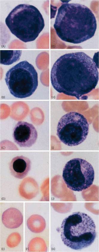
■ FIGURE 3-3 Microscopically recognizable stages of erythroid and granulocytic maturation: (A) proerythroblast; (B) basophilic erythroblast; (C) polychromatophilic erythroblast; (D) normoblast; (E) polychromatophilic erythrocyte; (F) erythrocyte (mature); (G) myeloblast; (H).promyelocyte; (I) neutrophilic myelocyte; (J) neutrophilic metamyelocyte; (K) band neutrophil. (From Cormack DH. Essential Histology. 2nd edn. Baltimore, MD:,Lippincott Williams & Wilkins, 2001.)
Lifespan and Numbers
After their development, leukocytes are circulated in the blood until the time (relatively short) they leave the circulation to perform their extravascular function. Granulocytes can be present in the blood for 6-20 hours and are constantly leaving.
Granulocyte time in the tissues varies considerably, but can be 2 or 3 days. Once granulocytes leave the blood, they do not normally return. They leave the body either from inflammatory sites or by way of the gastrointestinal, urinary, respiratory, or reproductive tracts. These organs are normally lined with neutrophils, which help prevent entry of organisms or foreign particles. Monocytes have a circulation time of 24 hours or less, but can remain in the tissues for several months. Many monocytes become fixed macrophages in the sinusoids of the liver, spleen, bone marrow, and lymph nodes; in this way they continue to function in the blood and lymph.Lymphocytes recirculate repeatedly from the blood to the tissues, to the lymph, and back to the blood. The lymphocyte population consists of T cells and B cells. Their lifespan varies, depending on classification. Generally T cells are long-lived (100-200 days), B cells are short-lived (2-4 days), and memory T and B cells are very long-lived (years).
The circulating leukocytes are considerably less numerous than erythrocytes. The numbers range from 7,000 to 15,000 per microliter (μL) among the domestic animals (Table 3-1). To appreciate the volume from which the number is obtained, recall that a microliter (μL) is one-millionth of a liter, whereas a milliliter (mL) is one-thousandth of a liter. Accordingly, there are 1,000 μL in 1 mL. The percentage distribution of the various types of leukocytes is not the same among the domestic species. There is a higher percentage of lymphocytes than neutrophils among the cloven-hoofed animals (pig, cow, sheep, goat). The reverse (higher percentage of neutrophils than lymphocytes) is true for the horse, dog, and cat.
TABLE 3-1 TOTAL LEUKOCYTES PER MICROLITER OF BLOOD AND PERCENTAGE O EACH LEUKOCYTE
| TOTAL LEUKOCYTE COUNT OF EACH LEUKOCYTE(RANGE; NO.∕μL) | PERCENTAGE OF EACH LEUKOCYTE | |||||
| SPECIES | NEUTROPHIL | LYMPHOCYTE | MONOCYTE | EOSINOPHIL | BA! | |
| Pig | ||||||
| .1 day | 10,000-12,000 | 70 | 20 | 5-6 | 2-5 | |
| ,1 week | 10,000-12,000 | 50 | 40 | 5-6 | 2-5 | |
| ,2 weeks | 10,000-12,000 | 40 | 50 | 5-6 | 2-5 | |
| ,6 weeks and older | 15,000-22,000 | 30-35 | bgcolor=white>55-605-6 | 2-5 | ||
| Horse | 8,000-11,000 | 50-60 | 30-40 | 5-6 | 2-5 | |
| Cow | 7,000-10,000 | 25-30 | 60-65 | 5 | 2-5 | |
| Sheep | 7,000-10,000 | 25-30 | 60-65 | 5 | 2-5 | |
| Goat | 8,000-12,000 | 35-40 | 50-55 | 5 | 2-5 | |
| Dog | 9,000-13,000 | 65-70 | 20-25 | 5 | 2-5 | |
| Cat | 10,000-15,000 | 55-60 | 30-35 | 5 | 2-5 | |
| Chicken | 20,000-30,000 | 25-30 | 55-60 | 10 | 3-8 | |
From Reece WO, Swenson MJ.
The composition and functions of blood. In: Reece WO, ed. Dukes’ Physiology of Domestic Animals. 13th edn. Ames, IA: Wiley-Blackwell, 2015.
Function
As a group, the WBCs serve as a defense mechanism against bacterial, viral, and parasitic infections and proteins foreign to the body. Each of the WBCs has a specific role in this broad function.
Neutrophils
The cell membranes of certain cells can engulf particulate matter (e.g., bacteria, cells, degenerating tissue) and extracellular fluid and bring them into their cytoplasm. The ingestion of particulate matter is known as phagocytosis, the ingestion of extracellular fluid is pinocytosis, and both are forms of endocytosis.
Neutrophils have two types of granules in their cytoplasm. Azurophilic granules are the lysosomes of the neutrophil and supply enzymes to digest ingested bacteria, viruses, and cellular debris. The other granules produce hydrogen peroxide, a bactericidal substance that is potentiated (made more active) by peroxidase, one of the lysosomal enzymes.
Substances within specific granules include collagenase and an iron-binding protein called lactoferrin. Lactoferrin has a very high affinity for ferric iron and can deprive phagocytized bacteria of the iron they need for further growth.
Neutrophils are highly phagocytic and this, coupled with their mobility, provides for an effective body defense mechanism. Their numbers increase rapidly during acute bacterial infections. The mechanism by which neutrophils move from the blood to an inflammatory site is described as follows (Figure 3-4)
1. Degenerative products of inflamed tissue or bacterial cells can be chemotactic (chemically attracting) and diffuse through interstitial spaces to capillaries and venules.
2. Chemotactic substances increase porosity of these vessels and also provide for adhesion of neutrophils to endothelium (margination).
3. Neutrophils squeeze through endothelial openings (diapedesis).
4. Neutrophils proceed to inflammatory sites by ameboid movement.
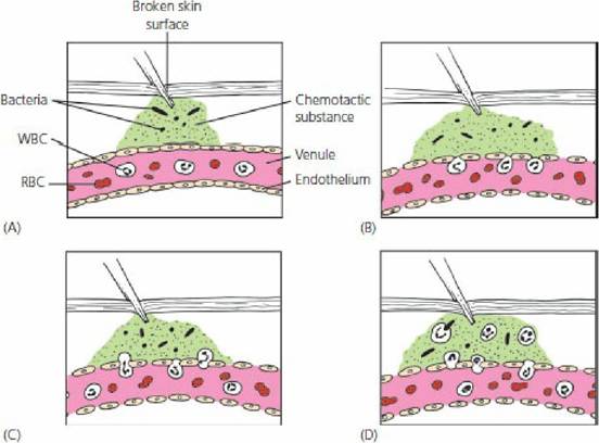
■ FIGURE 3-4 Mechanisms by which neutrophils are attracted to sites of injury. A. Tissue injury and introduction of bacteria causes diffusion of a chemotactic substance to capillaries and venules. B. Chemotactic substance increases endothelial porosity and adhesion of neutrophils to endothelium. C. By a process known as diapedesis, the adhered neutrophils squeeze through endothelial pores. D. Neutrophils proceed to an injury site by amoeboid movement and phagocytize bacteria and other debris. WBC, white blood cell; RBC, red blood cell.
This mechanism probably applies to the other leukocytes as well. When the neutrophils arrive at the inflamed site, they phagocytize bacteria and cell debris. The neutrophil lifespan is relatively short; dead neutrophils and their liquid is known as pus. The accumulation of pus within a connective tissue capsule is known as an abscess.
Monocytes
Monocytes are usually the largest leukocyte seen on a stained blood film. They occur in normal blood to only a limited extent. Compared with other leukocytes, they have a copious cytoplasm. Circulating monocytes phagocytize bacteria, viruses, and antigen-antibody complexes from the bloodstream. However, their circulatory phagocytic function is not as pronounced as that which occurs in the tissues. The movement of neutrophils from capillaries and venules is accompanied by similar margination and diapedesis of monocytes. On entering the tissues, monocytes are transformed into macrophages (large phagocytic cells) and initially participate in the phagocytosis of bacterial cells. Macrophages kill phagocytized microbes by their acidic pH, bacteriostatic proteins, and degradative enzymes. They also produce hydrogen peroxide in greater quantity than neutrophils. Macrophages eventually predominate at the inflammatory site because of their longer lifespan. Also, they are attracted to some organisms that neutrophils ignore and they phagocytize the cellular debris that remains when inflammation subsides.
The enzyme systems of monocytes are designed to degrade engulfed tissue debris from chronic inflammatory reactions and monocyte numbers increase in chronic infections. They are especially valuable in the defense against longterm inflammation because of their larger size and longer lifespan. Lysosomes within the cytoplasm of neutrophils and monocytes help in the digestion of the phagocytized materials.Monocytes are the cells that comprise the mononuclear phagocytic system (MPS). The MPS was formerly known as the reticuloendothelial system. Its cells are either monocytes (intravascular) or are derived from monocytes (extravascular). The cells are mobile (macrophages) or become fixed in position (e.g., Kupffer cells in the liver sinusoids and others in the spleen and lymph nodes). The fixed cells are also phagocytic.
Eosinophils
On a stained blood film, eosinophils can be seen to have cytoplasmic granules that,are red or reddish-orange (eosinophilic, see Figure 3-2). These are about the same size as neutrophils. The granules contain several enzymes (e.g., histaminase) that dampen and terminate local inflammatory reactions of allergic origin. Eosinophils become more numerous in certain types of parasitisms. The parasitic forms are opsonized (attacked by antibodies) and the eosinophils discharge their granular contents on to the surface of the opsonized parasite, inflicting lethal damage.
In Cushing’s disease there is oversecretion of adrenocorticosteroid hormones (see Chapter 6). When cortisol (an adrenocorticosteroid) is injected, this condition is simulated and the number of circulating eosinophils decreases. Cortisol reduces the eosinophil count by enhancing eosinophil diapedesis and by diminishing the release of eosinophils from the bone marrow. Cortisol production increases during stress and lowered eosinophil blood counts have been associated with stress.
Basophils
Basophils of the blood are somewhat similar to the mast cells that are present in the interstitial spaces outside the capillaries. They seem to lack phagocytic power. Basophil granules contain histamine, bradykinin, serotonin, and lysosomal enzymes, substances that initiate an inflammatory response. Basophils and mast cells have receptors on their cell membranes for immunoglobulin E (IgE) antibodies (those associated with allergies). When the antibody on the cell membrane contacts its antigen, the basophil ruptures, releasing its granular contents, and the local vascular and tissue reactions of allergies are manifested. Basophils are rare in normal blood and their distribution in blood is usually considered to be less than 1%.
Basophils enhance allergic reactions, whereas eosinophils tend to dampen them. There is a balance between their functions in that inflammatory reactions proceed quickly via basophils and then are modified via eosinophils so that overreaction does not occur.
Lymphocytes
Lymphocytes can be classified morphologically as small or large (see Figure 3-2). It is believed that large lymphocytes represent immature forms, whereas small lymphocytes represent more mature forms. Lymphocytes are involved in immune responses, and on this basis are classified as T cells or B cells. Both T and B cells are derived from hematopoietic stem cells (lymphoblasts) that differentiate to form lymphocytes. In mammals, shortly before or after birth, the site of early processing and differentiation of the stem cells for T lymphocytes is the thymus gland; for B lymphocytes, the sites are the fetal liver, spleen, and bone marrow. T cells are involved in cell- mediated immunity, which involves the formation of large numbers of lymphocytes to destroy foreign substances (antigens). The three different types of T cells are cytotoxic T cells, helper T cells, and memory T cells. Cytotoxic T cells are sometimes called killer cells. T-cell receptors bind to specific antigens and cytotoxic substances are released into the foreign cell (e.g., bacteria, viruses, tissue cells) (Figure 3-5).
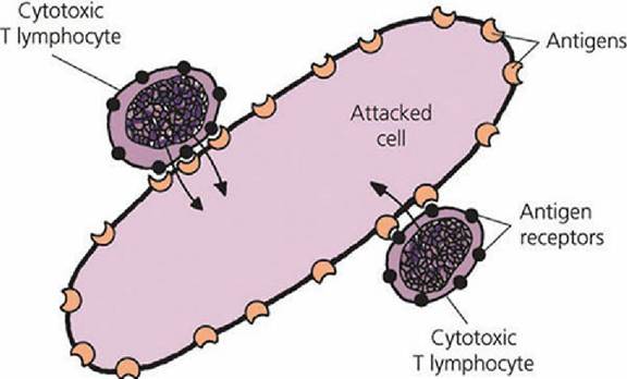
■ FIGURE 3-5 Mechanism by which sensitized cytotoxic T lymphocytes destroy a foreign cell. The attacked cell is killed by the release of cytotoxic and digestive enzymes from the T lymphocytes directly into the cytoplasm of the attacked cell. The T lymphocytes can proceed to other cells after their attack on a cell.
Cytotoxic cells also attack cells of transplanted organs. Because cancer cells generate unique antigens when they become cancerous, cytotoxic T cells recognize the cancerous cells as foreign to the body and attack them. Helper T cells are the most numerous of the T cells. When helper T cells are activated, they assist in the activation of cytotoxic T cells and of B cells. Antigens ordinarily activate the cytotoxic.T cells and B cells, but activation is more intense when assisted by helper T cells. Memory T cells are long-lived and respond to the same antigen when exposed at a later date.
B lymphocytes were first discovered in birds, and early processing and differentiation was found to occur in the bursa of Fabricius, from which the name was derived - B for bursa (see Chapter 12, Avian Digestion). After exposure to an antigen, activated B cells proliferate and transform into plasma cells and a smaller number of memory cells. The memory B cells have a function similar to memory T cells and are readily converted to effector cells by a later encounter with the same antigen. B cells do not attack foreign substances directly, but instead the plasma cells produce large quantities of antibodies (globulin molecules called immunoglobulins) that inactivate the foreign substance. This type of immunity is known as humoral immunity. Antibodies can produce inactivation by causing agglutination, precipitation, neutralization (antibodies cover toxic sites), or lysis (rupture of the cell). Agglutination and precipitation reactions are shown in Figure 3-6.
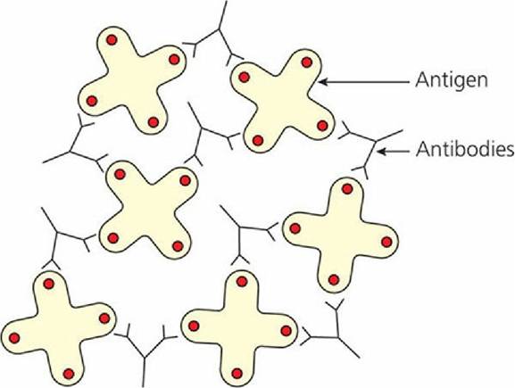
■ FIGURE 3-6 Antigen-antibody agglutination and precipitation. Antigens (molecules or cells) are grouped with other antigens by bivalent (two binding sites) antibodies. This causes them to agglutinate or precipitate. (Adapted from Hall JE. Guyton and Hall Textbook of Medical Physiology 12th edn. Philadelphia, PA: Saunders Elsevier, 2011).
A more common humoral method of immunity is represented by the complement system, which is composed of a number of enzyme precursors that are activated successively. From a small beginning, a large reaction occurs. Examples of complement reactions include (1) opsonization, in which foreign substances are covered by antibodies and become vulnerable to phagocytosis by neutrophils and macrophages, and (2) chemotaxis, in which the complement product attracts neutrophils and macrophages into the local region of the antigenic agent.
Diagnostic Procedures
Diagnostic procedures related to WBCs include determination of their total number and distribution of the leukocyte types. The total number can be determined by dilution and subsequent counting, either manually in a hemacytometer or with an electronic counter. An increase in leukocyte numbers is called leukocytosis; this usually occurs in bacterial infections. A decrease in numbers is called leukopenia; this is usually associated with the early stages of viral infections. Leukemia is a cancer of WBCs and is characterized by leukocytosis. The determination of the percentage distribution of WBCs is known as a differential white blood,cell count. In this procedure a smear is made of a blood drop, which is subsequently stained. The cells are observed under a microscope and the different types are counted and classified until a total of 100 have been tallied. The relative number for each type is then estimated as the percentage distribution in the blood (see Figure 3-1). The absolute number of leukocytes is calculated after the total number and differential count have been determined. The absolute number refers to the number per microliter for each leukocyte type. Determination of the absolute number can prevent misinterpretation of the differential count. For example, the total WBC count for a normal cow might be 9,000/pL. The relative number could be 30% neutrophils and 60% lymphocytes, in which the absolute numbers would be 2,700/pL (0.3 ? 9,000) and 5,400/pL (0.60 ? 9000), respectively. If traumatic reticuloperitonitis (hardware disease) is present, this same cow might have a total WBC count of 27,000/pL and a differential count of 70% neutrophils and 20% lymphocytes. A first interpretation might be that a lymphopenia exists (60% lymphocytes decreased to 20%). However, further calculation shows that the absolute number of lymphocytes remains the same (27,000/pL ? 0.20 = 5,400/pL), whereas the absolute number of neutrophils increases (27,000/pL ? 0.70 = 18,900/pL). The neutrophil increase would indicate inflammation. Refer to Appendix A, where normal absolute values (range and average) are presented for the domestic animal species.
■