External Ear
The external ear consists of the external auditory canal and its cartilaginous extension, the auricle (pinna). The auricle, sometimes known as the ear leather to dog fanciers, is shaped like a lopsided funnel, with a small cutaneous pouch on the caudal border a short distance above the ear opening (Figs.
11.40 and 11.41). There is a wide diversity in the shape, size, and posture (erect or folded) of dog ears. Most cats have erect auricles, but an exception is the Scottish Fold cat, in which the most distal portion of the auricle bends rostroventrally beginning at 3 to 4 weeks of age.The basis of the auricle is a plate of fibroelastic cartilage that is covered by subcutaneous tissue and skin. The skin on the inner (concave) surface adheres more firmly to the cartilage than that on the outer part.
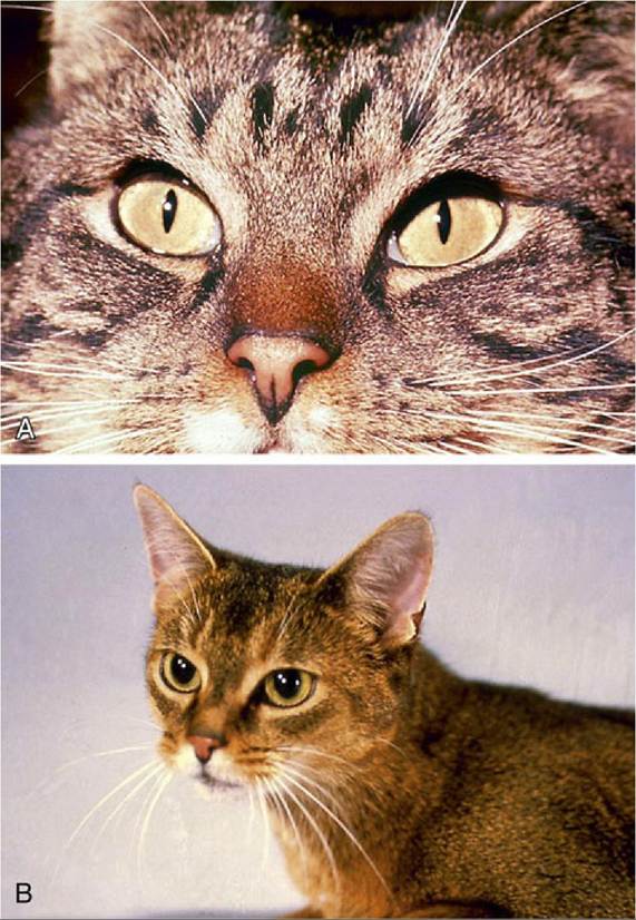
FIG. 11.38 (A) Slit form of constricted feline pupil. (B) Round form of dilated feline pupil.
The features of the auricular cartilage provide important surgical landmarks known as the helix, antihelix, tragus, antitragus, and scapha (see Fig. 11.41). The tragus, separated from the more caudal antitragus by the intertragic notch, forms the lateral rim of the ear canal opening. Both consist of rolled-up articular cartilage that supports the external ear opening. The antitragus forms the caudal part of the ear opening and ascends toward the end of the lateral side.
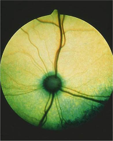
FIG. 11.39 Fundus of eye in a cat.

FIG. 11.40 Erect posture of the external ears.
The proximal part of the auricular cartilage is rolled to form a partial tube called the concha, which serves as the enlarged entry of the auditory canal.
This first part of the canal connects to the short anular cartilage, which terminates in a short osseous external canal. The ear canal is firstdirected ventrally (auricular cartilage) before turning medially to form the horizontal canal (portion of the auricular and anular cartilages), which is surrounded and supported by the temporal bone. This course hampers passage of the straight otoscope for examination of the proximal part of the canal and the eardrum. The examiner must straighten the canal by pulling the ear first caudally and then ventrally as the otoscope is advanced (Fig. 11.42). The canal is about 7 cm long.
FIG. 11.42
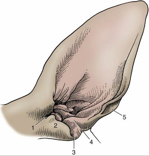
FIG. 11.41 Left canine ear, shaved. 1, Pretragic notch; 2, tragus; 3, intertragic notch; 4, antitragus; 5, cutaneous pouch.
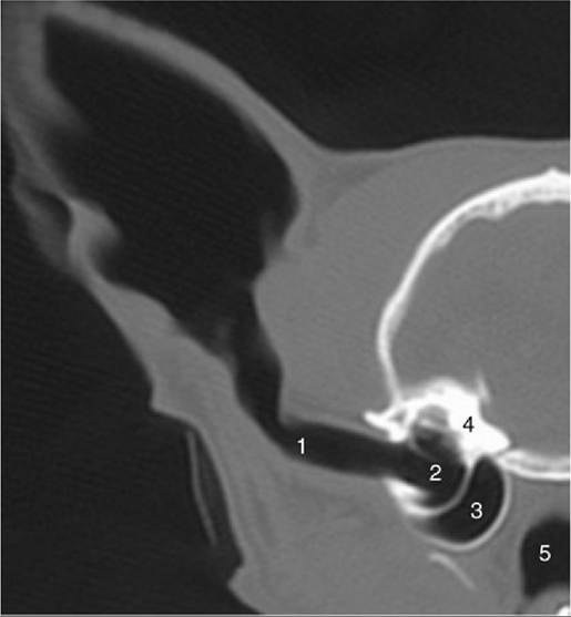
Transverse computed tomography scan (bone window) of half of a feline head showing ear
canal and middle ear. 1, Ear canal; 2, tympanic cavity; 3, tympanic bulla; 4, petrous temporal bone; 5, nasopharynx.
The horizontal ear canal ends at the eardrum. The tympanic membrane consists of an outer epithelial layer, which is a continuation of the skin of the external auditory canal, an inner mucosal layer, and a fibrous layer in between. The tympanic membrane is thin, slightly oval, semitransparent, and concave owing to traction on its medial side by the tensor tympani muscle (Fig. 11.43). The tympanic membrane consists of a small upper portion, the pars flaccida, and a large lower portion, the pars tensa (thin, tough, and glistening). The outline of the manubrium of the malleus is clearly visible.
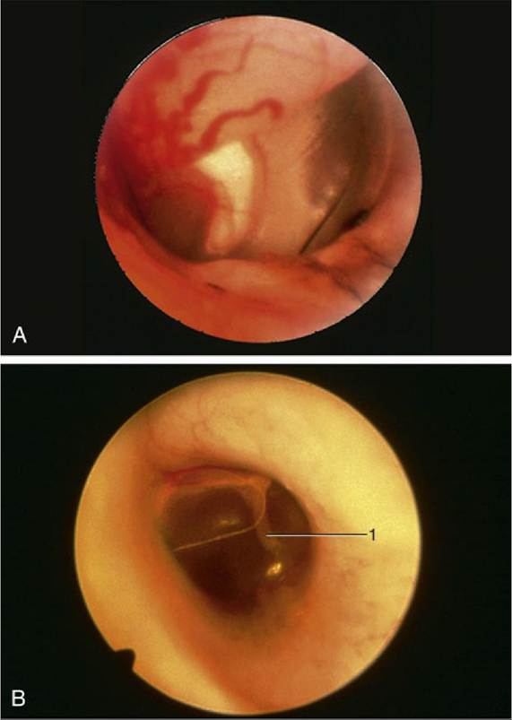
FIG. 11.43 (A) Otoscopic view of eardrum showing handle of malleus. (B) Otoscopic view of the eardrum (cat). 1, Malleus.
The auricular skin continues as the lining of the auditory canal. This skin is thin, and its lateral part possesses both ceruminous and sebaceous glands. It generally contains only a few hairs, but in some breeds (Poodles) hair is abundant. The skin of the bony part of the ear canal, which is much thinner than that of the cartilaginous portion, is continuous with the epithelial layer of the tympanic membrane. There are no glands or hair follicles here, where because of its thinness, the skin is more sensitive to trauma.
The base of the auricle and the ear canal are related laterally and ventrally to the parotid gland. The facial nerve crosses the ventral surface of the canal deep to the gland before breaking into the auriculopalpebral nerve and the two buccal branches. The former passes dorsally in front of the ear with the superficial temporal vessels. This stretch of the facial nerve also detaches a caudal auricular nerve and a branch to the middle ear. The sensory innervation is provided by the trigeminal, glossopharyngeal, vagus, and second cervical nerves. The innervation of the muscles of the external ear is by the facial nerve.
The veins of the area join the maxillary vein, which descends toward the mandibular gland from its formation by substantial caudal and cranial auricular and superficial temporal veins that may pass through the parotid gland (Fig. 11.44).
The arteries lie more deeply. The external carotid, having detached the caudal auricular artery to the convex surface of the auricle, ends rostroventral to the ear canal by dividing into maxillary and superficial temporal arteries. The latter, with the like-named vein, lies deep to the parotid gland close to the rostral surface of the ear canal.
The caudal auricular artery branches in the convex outer surface of the auricle and sends finer branches to the skin over the concave surface through small holes in the cartilage. Vigorous and repeated head shaking or scratching, in most instances elicited by parasites or infection of the ear canal, may injure the vessels and cause hematomas by rupture of the penetrating small branches. Because such a hematoma is lined by cartilage on both sides, splitting of the auricular cartilage also takes place. Once begun, the bleeding between the cartilages continues until the internal pressure equals the pressure in the feeder arteries.