Middle and Inner Ear
The middle and inner ears show few special features of importance. The auditory tubes are narrow and open on the dorsolateral wall of the nasopharynx, level with the landmark provided by the hamulus of the pterygoid bone, which is palpable through the mouth caudomedial to the last cheek tooth in the dog.
The tympanic bullae are large, hemispherical, and, except for a serrated septum in their rostral halves, undivided (see Fig. 11.23).In the cat, an incomplete bony septum bullae subdivides the middle ear into a small dorsolateral and a large ventromedial compartment. The two compartments communicate with each other through an opening at the caudodorsal margin of the septum near the cochlear window.
The middle ear infections (otitis media) may be drained into the nasopharynx through the bulla, which can be palpated through the oropharynx and soft palate, caudal to the hamulus. The inflated tympanic bulla of the cat is also easily found on palpation through the skin, between the wing of the atlas and the zygomatic arch.
The bulla can be approached surgically from the ventral side, with the use of the medial border of the rostral digastric muscle, the mylohyoid muscle, and the stylohyoid and tympanohyoid cartilages of the hyoid apparatus as landmarks; care should be taken to avoid damage to the nerves of the pharyngeal plexus (Fig. 11.44/12) and the vascular supply of the mandibular lymph node.
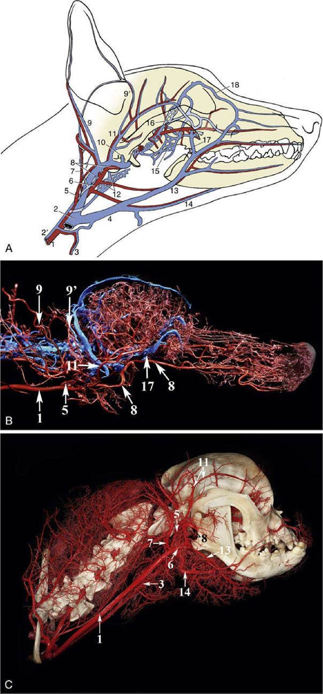
FIG. 11.44 The major arteries (red) and veins (blue) of the canine head. Schematic (A), corrosion cast without head (B) and with head (C). The ramus of the mandible has been removed. 1, Common carotid; 2, external jugular; 2', internal jugular; 3, cranial thyroid; 4, linguofacial; 5, internal carotid; 6, external carotid;
7, occipital; 8, maxillary; 9 and 9l, caudal and rostral auricular; 10, dorsal emissary; 11, superficial temporal; 12, ventral emissary and pharyngeal plexus; 13, facial; 14, lingual; 15, pterygoid plexus; 16, ophthalmic plexus; 17, deep facial; 18, angularis oculi.
Several nerves pass through the middle ear, but only two are of clinical significance. The facial nerve travels in the facial canal of the petrous temporal bone and detaches a branch, the chorda tympani, that enters the cavity of the middle ear.
Postganglionic fibers of the cranial cervical ganglion, located just behind the tympanic bulla, participate in a plexus within the middle ear. The resulting dysfunction is Horner syndrome, a complication of otitis media. The signs are miosis and retraction of the globe, which causes protrusion of the third eyelid, and narrowing of the palpebral fissure. The syndrome usually disappears spontaneously in about 3 months.