Eyeball position and movement
Key points
■ Seven extraocular muscles control eyeball position and movement; they are specifically innervated by cranial nerves III, IV and VI.
■ Head position and movement, sensed by the vestibular apparatus (CN VIII), stimulates cranial nerves III, IV and VI causing reflex positioning of the eyes - the vestibulo-ocular reflex (see Fig.
13.7).The eyes move in synchrony with each other, but different muscles for each eye are often activated to achieve conjugal eye movement. For example, moving the eyes to the left requires activation of the left lateral rectus muscle (left CN VI) and the right medial rectus muscle (right CN III). Dysfunction of CNN III, IV and VI results in strabismus (Figs. 10.6, 10.7).

Fig. 10.6 Extraocular muscles and strabismus. (A) Normal eyeball position, (B) ventrolateral strabismus (CN III dysfunction), (C) extorsional strabismus (CN IV dysfunction), (D) medial strabismus (CN VI dysfunction).
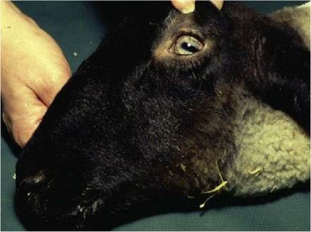
Fig. 10.7 Polioencephalomalacia showing strabismus secondary to paresis of the trochlear nerves; normally sheep have pupils that are horizontal, lined up with the medial and lateral canthi photo
(photo courtesy of Dr Phil Scott, University of Edinburgh).
Oculomotor nucleus (CN III)
The location of the oculomotor nucleus is in the ventral aspect of the periaqueductal grey matter in the midbrain. The efferent fibres pass to the ventral surface of the midbrain and form CN III (see Figs. A3, A18, A31). The oculomotor nerve supplies the levator palpabrae superioris muscle for elevation of the upper eyelid and extraocular muscles such as the dorsal, medial and ventral recti, and ventral oblique muscles.
Lesions cause ptosis (drooping of the upper eyelid) and ventrolateral strabismus.
The parasympathetic nucleus of CN III and oculomotor nucleus are located adjacent to each other and therefore lesions of that area usually involve both components. Damage to the parasympathetic portion will result in mydriasis and loss of the pupillary light reflex (see Visual reflexes, and Chapter 12).Trochlear nucleus (CN IV)
The location of the trochlear nucleus is in the midbrain just caudal to the oculomotor nucleus, at the level of the caudal colliculi. The efferent fibres pass lateral and dorsal to the mesencephalic aqueduct to exit on the dorsal aspect of the brain stem. All fibres decussate just ventral to the cerebellum in a thin membrane forming (the rostral medullary velum) the roof of the fourth ventricle and supply the contralateral dorsal oblique extraocular muscle (see Figs. A5, A7, A19). Lesions cause strabismus with outward rotation of the dorsal aspect of the eye (extorsion).
Abducens nucleus (CN VI)
The location of the abducens nucleus is in the rostral medulla oblongata just dorsal to the trapezoid body. Its efferent fibres supply the lateral rectus and retractor bulbi muscles (see Fig. A3). Therefore lesions cause medial strabismus and failure to retract the globe during eyelid closure as would normally be observed during menace response testing (see Chapter 13).