Visual reflexes
Key points
■ Visual reflexes are integrated in the midbrain.
■ The pupillary light reflex is driven by light and results in reflex constriction of the pupils bilaterally.
The reflex uses input via CN II (optic) and output via parasympathetic fibres of CN III.■ The direct (ipsilateral) reflex is stronger than the indirect (contralateral) reflex.
■ Head turning in response to visual stimuli is initiated by the rostral colliculi in the midbrain tectum.
Visual reflexes include head turning in response to visual and auditory stimuli (see Auditory reflexes), and the pupillary light reflex. The stimuli are received and integrated in the midbrain, from which arise connections to other cranial nerve nuclei and the spinal cord.
The pupillary light reflex (PLR)
The majority of afferent fibres travelling in the optic nerves (CN II) decussate at the optic chiasm and most fibres continue to the visual cortex via the lateral geniculate nuclei. A minority of fibres (20% in the cat) peel off the optic tract and synapse in the pretectal nucleus, which is located just rostral to the midbrain tectum between the rostral colliculus and the thalamus. The majority of efferent fibres from the pretectal nucleus decussate again in the caudal commissure, thereby projecting ultimately to the parasympathetic nucleus of III that is ipsilateral to the eye that was stimulated. However, because decussation is incomplete at both the optic chiasm and caudal commissure, a unilateral input will stimulate the parasympathetic nuclei of III bilaterally. Efferent parasympathetic fibres of III travel with the somatic fibres of the oculomotor nerve (CN III) to the orbit. The parasympathetic fibres synapse in the ciliary ganglion just caudal to the eye and postsynaptic fibres innervate the constrictor muscles of the iris and smooth muscle of the upper eyelid.
Because of various decussations, light stimuli cause reflex constriction of the ipsilateral pupil (direct reflex) and the contralateral pupil (indirect reflex). Since in domestic mammals the majority of fibres in the pupillary light reflex pathway cross in the optic nerve chiasm and the majority cross back again in the caudal commissure, the direct reflex is stronger than the indirect PLR (Figs. 10.8 and 10.9). In humans 50% of fibres cross each time, so there is no difference in the strength of pupillary constriction between the direct and indirect PLR. Conversely, in animals such as birds, in which there is complete decussation at the optic chiasm, the indirect reflex does not occur.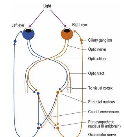
Fig. 10.8 Pupillary light reflex pathway.
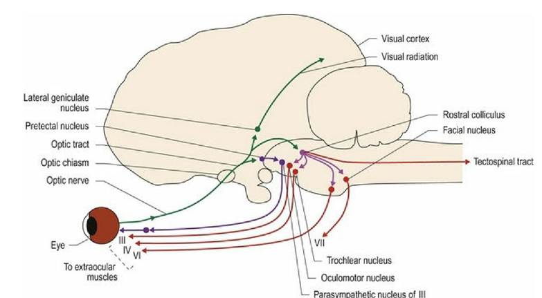
Fig. 10.9 Pathways involved in visual reflexes. Green = vision and visual processing, violet = interneuronal connections, purple = parasympathetic connections for pupillary light reflex, red = UMN and LMNs for eye, eyelid and head movement.
Note that the pupillary light reflex is a true reflex, not a response. It is incorrect to call it the pupillary light response. By comparison, the menace response is a learned function and it is incorrect to call that the menace reflex.
The midbrain comprises the dorsal tectum associated with primarily sensory function and the basal tegmentum associated with motor function. The paired rostral and caudal colliculi form the tectum (Figs. 10.11, A5, A7, A18-20, A31). The rostral colliculi are associated with visual reflexes; as such they are virtually non-existent in blind species such as the blind mole rat. (For the caudal colliculi function in auditory reflexes - see next section.) The rostral colliculi also receive input from the visual cortex and are important in vision. Output from the colliculi (tectum) form the tectonuclear tracts that pass into the brainstem providing UMN input to LMNs of CNN nuclei (CNN III, IV, VI).
Tectal output also forms the tectospinal tract influencing LMNs supplying the cervical muscles. Thus visual and auditory stimuli can cause reflex turning of the eyes or head, to allow the animal to focus on the stimulus (Fig. 10.9). The tectonuclear tract also connects to CN VII resulting in the protective dazzle reflex, in which bright light shone into the eye causes partial closure of the eyelids.Fig. 10.11
interthalamic adhesion.
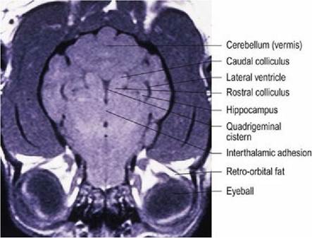
Dog brain, horizontal plane, T1-weighted magnetic resonance image at the level of the
In higher vertebrates, the colliculi are overshadowed by the occipital lobes, but in some fish, rodents and birds the optic tectum may be more important and is referred to as the optic lobe (Fig. 10.10). In these animals the primary input to the tectum is visual, however, it may also receive auditory, somatosensory and even electroreceptive stimuli. All inputs contribute to spatial mapping of the environment. With increasing development of the cerebral cortex and its connections, the physiological significance of the colliculi decreases.
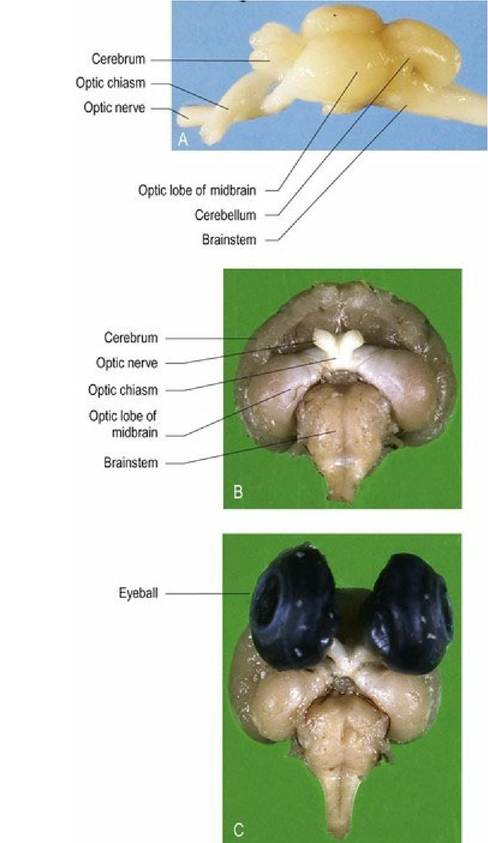
Fig. 10.10 Comparative brain anatomy. (A) Trout fish brain, dorsolateral aspect and (B, C) zebra finch brain, ventral aspect. Both species have well-developed optic lobes in the midbrain. Note that the optic
nerves of the trout brain are completely separate and have 100% cross over at the optic chiasm. (C) The size of the eyes explains the prominent optic nerves and lobe. Note: brains are not to scale with each other.
Each colliculus has many neural links through which visual and auditory functions are integrated. They are connected across the midline by a commissure. The rostral and caudal colliculi are connected rostrally to the lateral and medial geniculate nuclei, respectively, and to the pretectum and the thalamus. Caudally, there are bilateral projections via the tectoreticular, tectonuclear and tectospinal tracts.
The rostral colliculi, pretectum and the lateral geniculate nuclei are associated with visual reflexes such as head and eye turning in response to visual stimuli, coordinating eye movements and the pupillary light reflex. The function of the caudal colliculi is covered in the section on auditory reflexes.