FASCIA AND FAT
The connective tissue that separates and surrounds the more obviously important structures is generically known as fascia, a term of rather elastic usage; many of its larger accumulations, particularly those of a sheetlike nature, have specific names.
This tissue frequently receives scant notice, which is unwise, as it has significant functions to perform. Moreover, fascia is encountered in surgery, when it is necessary to predict its nature and extent in different situations.The superficial fascia (subcutis) is a loose (areolar) tissue extensively spread below the skin of animals that possess a hairy coat. A similar tissue surrounds many deeper organs, and in both situations, the loose fascia allows neighboring structures to change in shape and to move easily against each other. Its looseness varies with the amount of fluid it contains and may provide an indication of ill health. The superficial fascia is one of the principal sites for the storage of fat. In naked species, the fat forms a continuous layer, the panniculus adiposus.
The deep fascia is generally organized into much tougher fibrous sheets. A layer beneath the superficial fascia extends over most of the body and fuses to bony prominences. In many places it detaches septa that penetrate between the muscles, enclosing them individually or in groups (Figure 1-8); sometimes the periosteum, the fibrous covering of the bones, participates in outlining the enclosures. This division into fascial or osteofascial compartments is very prominent in the forearm and leg and plays a part in the circulation, assisting the return of blood and lymph to the heart. Muscles thicken when they contract, and when they are contained within
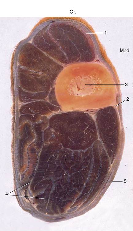
Figure 1-8 Osteofascial compartments in the forearm of a horse.
1, Superficial fascia; 2, cephalic vein; 3, radius; 4, septa of deep fascia enclosing individual muscles or groups of muscles; 5, deep fascia. (In transverse sections of the limbs, cranial [Cr.] and medial [Med.] are identified.)unyielding walls, they compress any other structures that share the space. If these are valved tubes (veins and lymphatic vessels), their contents are squeezed in one direction, toward the heart. Because of this, muscular paralysis or prolonged inactivity may lead to stasis of blood or lymph flow. Arteries and nerves whose functions would not be assisted by compression often travel in small tunnels within the septa.
More specific functions can be assigned to localized thickenings (e.g., retinacula: tethers) of deep fascia, which hold tendons in place and sometimes provide pulleys around which the tendons wind to change direction. Good examples are provided by the retinacula on the dorsal aspect of the hock and the palmar aspect of the digits (Figure 1-9/9).
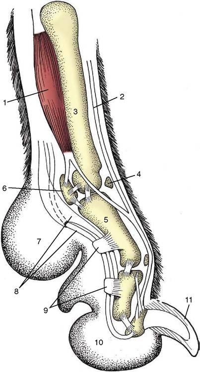
Figure 1-9 Axial section of a dog's paw; the metacarpal pad (7) is in contact with the ground during standing. 1, Interosseous; 2, extensor tendon; 3, metacarpal bone; 4, dorsal sesamoid bone; 5, proximal phalanx; 6, proximal sesamoid bone; 7, metacarpal pad; 8, flexor tendons; 9, retinacula; 10, digital pad; 11, claw.
Because dense fascia is relatively impermeable, it determines the direction taken by spreading fluids, such as pus that sometimes tracks below a fascial sheet before breaking through far from its source. This is one reason why some knowledge of the deep fascia is useful to the surgeon. Its toughness enables it to hold sutures securely while it also provides cleavage planes, which allow relatively bloodless access to deeper parts during surgery.
Most deposits of fat (adipose tissue) may be regarded primarily as food reserves. Small amounts of fat are widely distributed, but the bulk is contained in three or four places: in the superficial fascia (Figure 1-10/2); between and within muscles; below the peritoneum (the delicate membrane lining the abdominal cavity); and in the marrow cavities of long bones.
Subcutaneous fat deposits help mold the body contours and often show specific and gender differences in localization and development. Animals that are adapted to torrid habitats often develop localized depots (e.g., humped zebu cattle, camels, fat-tailed sheep), as a more even distribution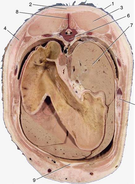
Figure 1-10 Transverse section of the back of a pig. 1, Skin; 2, fat (panniculus adiposus) associated with the superficial fascia; 3, back muscles; 4, cutaneous muscle enclosed within superficial fascia; 5, rib; 6, thoracic vertebra; 7, liver; 8, spinous process of vertebra; 9, additional fat deposited between muscles.
might interfere with heat loss to the environment. Some of the differences in the body form of men and women that become accentuated at puberty are produced by the deposition of fat in the breasts and over the hips and lower abdomen of females. In many male animals, much fat is deposited in the tissues of the dorsal part of the neck: the thickened crest of stallions is a good example.
Some fat deposits, like that enclosed within a fibrous lattice in the footpad of the dog, function as mechanical buffers (see Figure 1-9/7,10). Fat with a mechanical function is usually resistant to mobilization in starvation.
Differences in the chemical and physical nature of fat can be pronounced but may reflect diet as much as specific genetic factors. When the origin of a specimen is being determined, it is certainly often useful to know that the fat of horses and of Channel Island breeds of cattle is yellow, that of sheep hard and white, and that of pigs soft and grayish. It should also be remembered that fat at body temperature is softer (semifluid) than that exposed in a colder environment. Certain procedures—liposuction and lipofixation—employed by the cosmetic surgeon depend on this fortunate circumstance.
All these remarks refer to the common sort of fat. A second variety, brown fat, is of much more restricted distribution in time and place.
Brown fat differs in structure (Figure 1-11) and function as well as in color. In domestic species, it is especially found during the fetal and neonatal periods; in wild species, it is especially prominent in those that hibernate (Figure 1-12). The brown adipocyte contains numerous smaller droplets and a much higher number of mitochondria. It is richly vascularized. It provides both groups with a readily available source of heat, equally useful in newborn animals with imperfect thermoregulation and in hiber-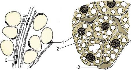
Figure 1-11 Fat cells of white (left) and brown (right) fat. In white fat a single large fat vacuole displaces the cytoplasm and the nucleus to the periphery of the cell. The small fat vacuoles are evenly distributed in the cells of brown fat. 1, Nuclei; 2, fat vacuoles; 3, capillaries.
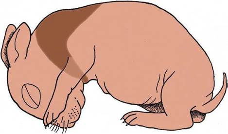
Figure 1-12 The distribution of brown fat in the newborn rabbit, concentrated around the neck and between the shoulder blades.
nators required to awaken rapidly from a deep winter sleep.
BONES
The primary functions of the skeleton are to support the body, to provide the system of levers used in locomotion, and to protect soft parts. Therefore, biomechanical factors are most important in shaping the bones and in determining their microscopic design. The major skeletal tissue, bone, has a secondary role in mineral homeostasis, supplying a reserve of calcium, phosphate, and other ions.
The Classification of Bones
Bones may be classified in various ways. A topographical classification recognizes a cranial skeleton (of the head) and a postcranial skeleton consisting of two divisions: the axial skeleton of the trunk and the appendicular skeleton of the limbs. A second classification based on ontogeny distinguishes the somatic skeleton, formed in the body wall, from the visceral skeleton, derived from the pharyngeal (branchial) arches.
A third system is also based on development and distinguishes parts preformed in cartilage (and later largely replaced by bone) from those that ossify directly in fibrous connective tissue. This classification reflects the phylogeny, as bones that develop in membrane are homologous with dermal bones of lower vertebrates.Individual bones are classified by shape according to a rather naive system (Figure 1-13). Long bones, which are typical of the limbs, are broadly cylindrical and are clearly adapted to perform as levers. It is perhaps more important to know that they develop from at least three centers of ossification: one for the shaft (diaphysis), and one for each extremity (epiphysis) (p. 72).
Short bones have no dimension that greatly exceeds the others. Many are grouped together at the carpus and
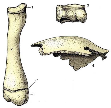
Figure 1-13 Long, short, and flat bones. 1, Proximal and distal epiphyses; 1', epiphysial cartilage; 2, diaphysis of a young dog's radius; 3, carpal bone of a horse; 4, parietal bone from the skull of a dog.
tarsus, where the multiplication of articulations provides for complex movements and may also diminish concussion. The majority of short bones develop from a single center of ossification; replication of centers generally indicates that the bone represents the fusion of elements distinct in ancestral forms.
Flat bones are expanded in two directions. The category includes the scapula, the bones of the pelvic girdle, and many of those of the skull. Their broad surfaces afford attachment to large muscle masses and protection to underlying soft parts.
The remaining bones are too irregular in form to be grouped in clearly defined categories. Neither flat nor irregular bones exhibit uniformity in development.
The Organization of a Long Bone
Many features of bone construction are conveniently approached through the examination of a longitudinal section of a long bone (Figure 1-14, A).
The form of the bone is determined by a sheath or cortex of solid (compact) bone that is composed of thin lamellae arranged mainly in series of concentric tubes about small central canals. Each such system is known as an osteone (Figure 1-14, B). The cortex is thick toward the middle of the shaft but thins as it flares toward each extremity, over which it continues as a crust. The external surface is smooth except where irregularities serve as the attachment sites for muscles or ligaments; these irregularities may be raised or depressed and, in both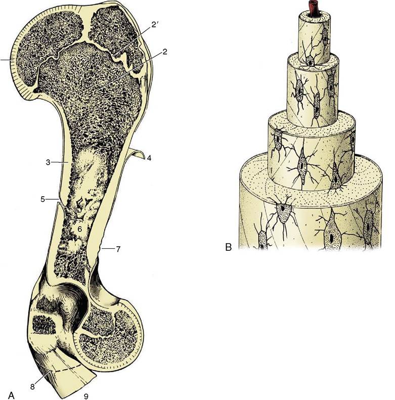
Figure 1-14 A, A long bone (bovine humerus) sectioned longitudinally. B, Osteone with central (haversian) canal. 1, Articular cartilage; 2, spongy bone; 2', epiphysial cartilage; 3, compact bone; 4, periosteum, partly reflected; 5, nutrient foramen; 6, marrow cavity; 7, roughened area for attachment of muscle or ligament; 8, distal extent of medial epicondyle; 9, tendons of origin of carpal and digital flexors.
cases, permit a concentration of the attachment. These features are generally most pronounced in larger, older males. They are given a variety of descriptive names of conventional significance; most elevations are known as lines, crests, tubercles, tuberosities, or spines; most depressions, are known as fossae or grooves (sulci).
The inner surface of the shaft bounds a central medullary (marrow) cavity and is rough; the irregularities are low, indiscriminate, and without apparent significance.
The extremities are occupied by cancellous or spongy bone, which forms a three-dimensional lattice of interlacing spicules, plates, and tubes of varying density.
The medullary cavity and the interstitial spaces of the spongy bone are occupied by bone marrow, which occurs in two intergrading forms. Red bone marrow is a richly vascularized, gelatinous tissue with hemopoietic properties; it produces the red and granular white corpuscles of the blood. Although all marrow is of this type in the young animal, most is later infiltrated with fat and converted into waxy yellow marrow whose hemopoietic potential is dormant. It is the marrow in the larger spaces that first becomes inactive, then that of the spongy bone of the distal limb bones, until finally active marrow is confined to the proximal extremities of the humerus and femur, the bones of the limb girdles, and those of the axial skeleton. The chronology of these events for domestic animals is uncertain.
The parts that articulate with neighboring bones are smooth. These articular surfaces are more extensive than the areas in contact in any position of the joint and provide a range of movement. They are clothed in hyaline articular cartilage. The cartilage is not uniform in structure; it is calcified in its deepest layer, which is firmly attached to the underlying cortex, and becomes fibrous toward the periphery, where it blends with the periosteum and joint capsule.
A tough fibrous membrane, the periosteum, ensheathes the remainder of the outer surface, from which it can be readily stripped, except where it is penetrated by tendons and ligaments proceeding to anchor in the compacta. Its appearance is rather misleading because the deeper layer is cellular and, even in adults, retains the bone-forming capacity that it exercised during development (p. 72). This osteogenic function is reactivated in the healing of a fracture.
Bones have a generous blood supply, perhaps amounting to 5% to 10% of the cardiac output. Several sets of vessels exist; the so-called nutrient artery, though generally the largest single source, probably contributes less than do the others in the aggregate. The nutrient artery penetrates toward the middle of the shaft in a position that is fairly constant for each bone. It is usually directed toward one extremity, and the foramen through which it passes may simulate an oblique fracture when depicted in radiographs. The artery divides into two divergent branches within the marrow; these and the later divisions pursue very tortuous courses, which may have the purpose of reducing the pressure within the vessels of the delicate marrow (Figure 1-15). The smaller branches supply the sinusoids of the marrow tissue and also the arterioles and capillaries that permeate a system of tiny central channels (haversian canals) within the osteones of compact bone. A further supply to the cortex arises from the medullary sinusoids. Branches of the nutrient artery that reach the metaphysial region (the part of the shaft adjacent to the epiphysis) anastomose there with branches of metaphysial and epiphysial vessels that enter the bone toward its extremity. The central region of this part of the shaft probably relies mainly on the nutrient artery, whereas the peripheral part relies on metaphysial arteries. The anastomoses are of varying efficiency, but the collateral circulation is generally sufficient to allow a bone to survive deprivation of part of its usual supply when fractured. One technique (intramedullary pinning) employed in fracture repair is possibly even more damaging to the vessels than is the initial injury, and its success serves to emphasize the value of the anastomoses. Some authors have described an additional supply entering the cortex from numerous small periosteal arteries. The weight of opinion denies their presence in healthy young bones.
Figure 1-15 The blood supply of a long bone, schematic. The supply of the cortex is shown (enlarged) in the center. 1, Epiphysial arteries; 2, metaphysial arteries; 3f nutrient artery; 4, 4'f artery and vein of the bone marrow; 5, periosteal arteries; 5'f periosteal vein; 6, anastomosis between periosteal and bone marrow arteries; 7, capillaries of the cortex; 8, sinusoids in the bone marrow; 9, growth cartilage; 10, cortex.
The main drainage of the marrow is effected by large, thin-walled veins that accompany the major arteries and emerge through the nutrient, epiphysial, and metaphysial foramina. The capillaries within cortical tissue drain into venules within the periosteum. The normal cortical circulation is therefore centrifugal—from within outward. No lymphatic vessels are present within bone, although infections of bone may spread to the lymphatics that drain neighboring tissues.
One important difference is exhibited by the circulation in young growing bones. In these, the circulation within the epiphyses forms separate and independent compartments, as (with few exceptions) arteries do not penetrate the growth (epiphysial) cartilage.
Nerves accompany the larger vessels, and their branches are to be found within the central canals of the osteones. Some (vasomotor) fibers pass to the vessels, some are sensory to the bone tissues (especially the periosteum), and the destination of others remains unclear. It is no longer believed that nerves exert a trophic influence on bone.
Biomechanical Aspects
It has long been the convention to explain the tubular construction of long bones by drawing the comparison with a loaded beam of some stiff, homogeneous material supported at both ends (Figure 1-16). In this construction the tensile forces that tend to disrupt the material are concentrated toward the lower surface while the compressive forces that tend to crush and compact the material are concentrated toward the upper surface. These forces tend to neutralize each other along, and close to, the axis, and the material here is more or less redundant. It can be dispensed with or replaced by some weaker but lighter material, as in a
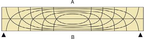
Figure 1-16 Pattern of compressive (A) and tensile (B) stress lines in a beam supported at both ends. The greatest stresses (closeness of lines) occur in the middle of the beam toward the surfaces.
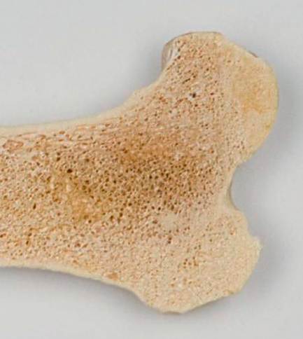
Figure 1-17 Proximal end of the humerus of a cow, sectioned sagittally, as an example of the architecture of spongy bone.
long bone. The analogy is not exact—for a start, bone is a composite material—but it is useful as a first approach. The diagram (see Figure 1-16) shows that the lines of principal compressive and tensile stress intersect in orthogonal fashion toward the extremities of the model; the spongy architecture of a bone closely mimics the theoretical pattern. Indeed, the pattern of trabecular bone has been described as the crystallization of the lines of stress, which is an attractive if faulty metaphor. Because the more detailed analysis of the spongy architecture (Figure 1-17) introduces matters that are both complicated and controversial, it is probably wiser to leave discussion to the specialist.
Compact bone is a plastic, composite material of considerable strength, capable of sustaining and recovering from considerable deformation. When bent, the lamellae and osteones of which compact bone is constructed first shear past each other; if bent too far, a crack appears at right angles to the line of shear and then quickly spreads to create a brittle fracture. Most fractures are caused by excessive bending, which stresses both aspects of the bone approximately equally. Since the side under tensile stress generally fails first, this indicates that compact bone is better able to resist compression. However, spongy bone is commonly crushed and impacted by compression.
Some Specialized Varieties of Bones
Bones are often found within tendons (rarely within ligaments) where they change direction over prominences that would expose them to excessive pressure and friction. These bones, known as sesamoid bones, form regular synovial joints with the major bones with which they are in contact. In addition to preventing tendon wear, a sesamoid bone displaces the tendon farther from the axis of the adjacent joint and so serves to increase the leverage exerted by the muscle. The best- known example is the patella (kneecap) that forms in the principal muscle that extends the stifle joint (the name given to the knee of quadrupeds) (see Figures 2-63 and 17-3). In the dog, smaller sesamoids also develop in muscles behind the stifle, in the tendons passing behind the metacarpophalangeal joints (at the bases of the digits), and in the extensor tendons within the digits (see Figure 1-9). The chief practical importance of these and other lesser sesamoids lies in the risk of their being wrongly identified as chip fractures when they are depicted in radiographs. In large animals, one or more additional sesamoids form dorsal to the deep flexor tendon shortly before its insertion on the distal phalanx (or phalanges). In the dog the reaction is limited to the development of a nubbin of cartilage in each branch of the tendon.
Although sesamoids are a device to protect tendons from injury, the major sesamoids develop in the embryo before movement is possible, and their origin must therefore be genetically determined. They do not reform after extirpation when the limb is immobilized but only if movement is allowed; this indicates that they can also develop in reaction to an appropriate stimulus in the lifetime of the animal.
Splanchnic bones develop in soft organs, remote from the rest of the skeleton. The most familiar, indeed the only significant, examples in veterinary anatomy are the os penis (and the female equivalent, os clitoridis) of the dog and cat and the ossa cordis found in the heart, especially in the hearts of ruminants.
Certain bones are excavated to contain air spaces. In mammals, these pneumatic bones are confined to the skull and contain the paranasal sinuses, which communicate with the nasal cavities. The sinuses principally develop after birth, when outgrowths of the nasal mucosa invade certain skull bones and replace the diploe, the spongy bone between the outer and inner layers (“tables”) of compacta. The separation of the tables can be very considerable and can lead to a remarkable postnatal remodeling of the skull, best exhibited by cattle and pigs. The postcranial skeleton of birds develops an extensive system of air-filled cavities in communication with the respiratory organs.