FUNCTIONAL ANATOMY OF FEMALE MAMMARY GLANDS
1. Note the relationship of alveoli, lobules, lobes, ducts, and lactiferous sinus (gland cistern, teat cistern).
2. What is the milk-secreting unit of the mammary gland? Is it part of the parenchyma or stroma?
3.
Do teat canal, papillary duct, and streak canal refer to the same thing?4. What seems to be the function of the rosette of Furstenberg?
5. What seems to be the function of the venous plexus in the teat wall?
6. What function is served by inward folds of the empty teat wall?
7. What function is served by the sphincter muscle that encircles the streak canal?
8. What function is served by the suspensory apparatus?
9. What is the venous circle of the mammary gland, and where is it located? What is the milk vein? What is the milk well?
0. What is the function of the myoepithelial cells?
The physiology of lactation among the domestic animal species is similar; however, anatomic differences exist related to outward appearance, location, and numbers of glands, teats, and teat openings. Because of their widespread use in milk production, the bovine female will be presented as the lactation model.
Mammary Gland of Cows
The mammary gland (udder) of the cow has an inguinal location with distinct right and left halves, and each half has a front and hindquarter (Figure 16-1). Each half is independent from its counterpart in regard to its blood and nerve supply, lymphatic drainage, and suspensory apparatus. A longitudinal furrow marks the ventral separation of the halves. The two quarters of each half are separate in regard to gland tissue and duct system. All of the milk from one teat is produced by the glandular tissue of that quarter. The parenchyma of the mammary gland refers to the epithelial or glandular tissues, as opposed to the stroma, which is the connective tissue framework of the mammary gland.
The milk-secreting unit of the mammary gland is the alveolus (Figure 16-2). A number of alveoli converge on lactiferous ducts that convey milk to a gland cistern, and finally to a teat cistern. The gland cisterns and teat cisterns are collectively known as lactiferous sinuses (see Figure 16-1). A number of alveoli grouped together and surrounded by a layer of connective tissue is known as a lobule. A larger connective tissue division surrounds a number of lobules to form a lobe. The secreting units of the mammary gland are thus divided into lobules and lobes.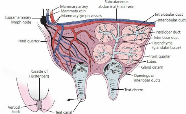
■ FIGURE 16-1 Sagittal section of the cow udder through the left half. The four circular areas in the front quarter are schematically shown to illustrate the organization of the glandular tissue and also the various orders of ducts. The lobes are distributed throughout the parenchyma. The lobes are further divided into lobules (not shown). The gland cistern and teat cistern for each quarter are collectively known as the lactiferous sinus. The teat canal magnification shows the vertical folds of the teat canal and also the rosette of Furstenberg at the upper end.
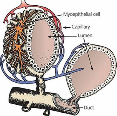
■ FIGURE 16-2 Alveolus surrounded by blood vessels and myoepithelial (contractile) cells. Several alveoli in a group form a lobule. Each alveolus converges on an intralobular duct. (From Larson BL. Biosynthesis and cellular secretion of milk. In: Larson BL, ed. Lactation. Ames, IA: Iowa State University Press, 1985.)
The Teat
The part of the mammary gland from which milk is extracted and suckled by the young is called the teat, with one teat for each quarter of the udder. A section of the cow’s teat is shown in Figure 16-3. The duct extending from the teat cistern to the teat orifice is the papillary duct (teat canal). The teat canal is more commonly known as the streak canal.
It is normally closed by a smooth muscle sphincter (reinforced by elastic tissue) that encircles the streak canal (Figure 16-4). Closure of the streak canal prevents leakage of the milk that accumulates within the lactiferous sinus. The mucosa of the streak canal is marked by vertical ridges that radiate upward from the steak canal’s internal opening into the teat cistern, forming the rosette of Furstenberg (see Figure 16-1 and Figure 16-4), which appears as folds of mucosa. The weight of milk in the lactiferous sinus exerts a downward thrust on the folds, thus covering the inner opening to the streak canal and assisting with retention of milk within the udder. External pressure and downward pull on the teat at milking causes the overlapping folds to be withdrawn so that milk can escape through the teat orifice. Inflammation or injury to the rosette can lead to excessive development, resulting in partial restriction or blockage of the streak canal. Furstenberg rosette epithelial cells contain keratin that function to trap bacteria that may invade the teat. As milk flows through the streak canal, the keratin peels off along with any trapped bacteria, thus removing bacteria from the teat. The wall of the empty teat cistern is characterized by numerous longitudinal and circular folds. When the teat is filled with milk, these folds are obliterated. The presence of the folds permits expansion of the teat wall without tension. The venous plexus of the teat wall (see Figure 16-3) constitutes a form of erectile tissue and becomes congested when the teat is stimulated.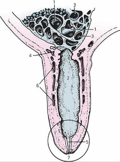
■ FIGURE 16-3 Sagittal section through a cow’s teat. 1, Gland cistern; 2, teat cistern (gland cisterns and teat cisterns are collectively known as lactiferous sinuses); 3, openings of interlobar ducts; 4, submucosal venous ring; 5, teat canal; 6, venous plexus in teat wall; 7, streak canal (circled area is shown in Figure 16-4).
(From Dyce KM, Sack WO, Wensing CJG. Textbook of Veterinary Anatomy. 3rd edn. Philadelphia. PA: WB Saunders, 2002.)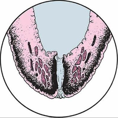
■ FIGURE 16-4 Section of the teat (circled area as shown in Figure 16-3) showing the smooth muscle encircling the teat canal (papillary duct). The thickened area above the teat canal, which would encircle it, is the rosette of Furstenberg. (From Dyce KM, Sack WO, Wensing CJG. Textbook of Veterinary Anatomy. 3rd edn. Philadelphia, PA: WB Saunders, 2002.)
The ease with which milk can be withdrawn from the teat is often determined,by the tightness of the sphincter that keeps the teat canal closed. A sphincter that is,not tight enough can allow milk to leak from the teat in the interval between milkings. A loose sphincter also predisposes to mastitis (inflammation of the mammary gland usually resulting from infection by microorganisms).
Suspensory Apparatus
Support from the longitudinal axis of the body is provided to the udder by the suspensory apparatus, which is composed of medial and lateral suspensory ligaments (Figure 16-5). The medial suspensory ligament is derived from the elastic fibers (connective tissue) that cover the abdominal wall. It passes down between the two halves of the udder and intimately covers the medial side of each half, passes around the front to about the middle of the cranial quarters, and passes around the back to about the middle of the caudal quarters. The lateral suspensory ligaments are composed of white, fibrous, connective tissue (with little elasticity) derived from the subpelvic tendon. The lateral ligaments cover the lateral side of each half and meet the medial suspensory ligament at the front and back of each half. A number of connective tissue extensions (laminae) are given off from both the medial and lateral suspensory ligaments to enter the mammary gland. The laminae divide each quarter into lobes and lobules.
Collectively, the laminae form the stroma (framework) of the mammary gland.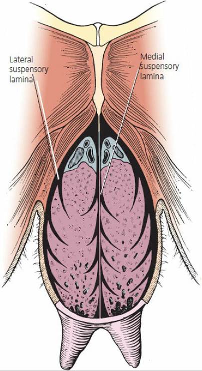
■ FIGURE 16-5 Suspensory apparatus of the cow. The udder is shown in transverse section through hindquarters. (From Frandson RD, Wilke WL, Fails AD. Anatomy and Physiology of Farm Animals. 7th edn. Ames, IA: Wiley-Blackwell, 2009.)
The function of the elastic fibers in the suspensory apparatus becomes apparent when the cow is mature and in production. The elastic fibers allow for absorption of the shock created when the cow walks and permit movement of the udder while the cow is lying down.
Blood Supply and Venous Drainage
The principal blood supply to each half of the mammary gland is the external pudendal artery (called the mammary artery in the cow) (see Figure 16-1). It passes through the inguinal canal and divides to supply the front and hindquarters on the same side as the artery. The external pudendal vein (mammary vein in the cow) collects blood from the cranial and caudal quarters of the respective side and returns blood through the inguinal ring to the caudal vena cava (Figure 16-6). The mammary veins are continuous cranially with the subcutaneous abdominal milk veins and caudally with anastomosing (joining) ventral labial veins so that a venous circle is formed at the base of the udder. The milk veins are relatively large, tortuous (winding) veins on the ventrolateral wall that disappear suddenly at a forward location (milk well) to enter the internal thoracic vein.
Some believe that blood can enter the venous circle from the milk veins (a reverse direction). As in other tissues, the interstitial fluid has auxiliary drainage by way of lymphatic vessels with lymph nodes along their length. The major lymph node on each side is the superficial inguinal (mammary or supramammary) lymph node, located near the inguinal ring above the caudal part of the base of the udder (see Figure 16-1).
Capillary networks are present, as in other tissues, and for the udder these surround the alveoli and ducts much as capillaries surround alveoli in the lungs (see Figure 16-3).
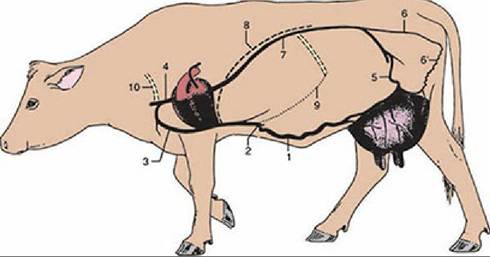
■ FIGURE 16-6 Venous drainage of the udder. 1, Subcutaneous abdominal (milk) vein; 2, milk well; 3, internal thoracic vein; 4, cranial vena cava; 5, external pudendal vein (mammary vein); 6, internal pudendal vein; 6', ventral labial vein; 7, caudal vena cava; 8, diaphragm; 9, costal arch; 10, first rib. (From Dyce KM, Sack WO, Wensing CJG. Textbook of Veterinary Anatomy. 4th edn. St Louis, MO: Saunders Elsevier, 2010.)
Myoepithelial Cells
The myoepithelial cells are contractile cells that surround the alveoli and ducts. Because of their location relative to the alveoli,.they have been called basket cells (see Figure 16-2). When contracted, they provide compression on the alveoli and ducts and hence cause milk to be directed toward the lactiferous sinus. They contract when the hormone oxytocin circulates and brings about milk letdown.
Mammary Glands of Other Animals
Pigs, Dogs, and Cats
The sow normally has seven pairs of mammary glands (range, four to nine). The teat of the sow has two teat canals, and each is continuous with its respective teat cistern, gland cistern, and associated ducts. In the bitch and queen, five pairs of mammary glands are most common, and each mammary gland has a mammary papilla, or nipple. The nipples have numerous fine openings (seven to 16) at their distal ends for the ducts of the glands. In the sow, bitch, and queen, the mammary glands are located in two rows parallel to the midline.
Sheep and Goats
The mammary gland has an inguinal location in both the ewe and doe. Each half has only one teat, one teat canal, one teat cistern, and one gland cistern. The sphincter muscles at the tips of the teats are poorly developed and closure is assisted by elastic connective tissue.
Horse
The horse has an inguinal location for its mammary gland and each half has only one teat. Each teat has two teat canals and two teat cisterns; each teat cistern is continuous with a gland cistern that has its own system of ducts and alveoli.