Functional Changes
It is often stated that bitches come in heat twice a year, in spring and autumn. In fact, three heats are not uncommon, although even in bitches with three heats, the greater part of the year is occupied by periods of anestrus.
Cats may even have four cycles in place of the usual two. The first heat occurs at the age of 6 to 9 months in bitches and at 6 to 12 months in young queens, depending on the season of their birth.The reproductive organs, quiescent during anestrus, develop rapidly in proestrus, when over a period of a week, a batch of follicles enlarges. The uterus now increases in length and in thickness, its endometrium proliferates, and the entire reproductive tract becomes hyperemic. A thickened, edematous vulva discharges the blood-tinged serous uterine secretion. Estrus also lasts about a week and can be distinguished from proestrus by the female's readiness to accept a male. The endometrial hypertrophy and hyperemia continue, but the discharge gradually becomes less bloodstained. Ovulation, which occurs about the second day of estrus, is succeeded by very rapid formation of corpora lutea, which may be mature by the end of estrus.* The separation of diestrus and metestrus is difficult to determine because there is often a period (2 to 8 weeks) of pseudopregnancy, during which the bitch exhibits the usual physical and behavioral signs of pregnancy even though fertilization has not occurred; pseudopregnancy can perhaps be likened to a greatly extended period of diestrus. The cervix is tightly closed during diestrus and metestrus, and secretions that would have been utilized for embryo nutrition then accumulate in amounts that may distend the uterus. Sometime infection of the uterus (pyometra) may necessitate hysterectomy.
In comparison with other domestic species, the bitch's vaginal epithelium responds in a more pronounced manner to changes in hormonal levels, and the vaginal smears provide evidence of the stage within the cycle.
Both cornified epithelial cells and erythrocytes are present in large numbers during proestrus. The cornified epithelial cells persist in estrus, but the erythrocytes gradually become fewer and leukocytes appear. The stages of the cycle are also reflected in the gross appearance of the vaginal lining, including that covering the dorsomedian fold. In proestrus the lining becomes edematous, and forms prominent soft folds. As estrogen levels drop rapidly during estrus, the vaginal wall becomes less edematous, and the lining wrinkles until about 4 days after ovulation, when the surface is said to resemble crepe paper. A few days later the mucosa becomes flat and patchy; with the desquamation of the cornified superficial layer of epithelium the blood vessels are able to shine through once more.Ova enter the uterus about the sixth day after ovulation. The fertilized ova implant after another 10 days. The initially established omphalovitelline (yolk sac) is later replaced by the definitive chorioallantoic placenta (Fig. 15.14/6). The placenta develops through the invasion of the endometrium by villi growing from a broad band of the chorion encircling the trunk of the fetus and is a continuation of the erosion that started in the nonvascular (chorioamniotic) regions and about the yolk sac attachment. The erosion leads to the interdigitation of thin plates of fetal tissue, and endometrial lamellae are reduced to little more than the maternal capillary endothelium (see Fig. 5.70E-H). The tissue barrier of this basically Chorioendothelial placenta is further reduced at the margins of the zonary band, where blood extravasated from maternal vessels directly bathes the fetal tissue. Hemoglobin breakdown in these marginal hematomas is responsible for the brilliant green pigmentation that contrasts with the deep red of the major part of the placenta (see Fig. 5.67A). In short, this type of placenta consists of three zones: a transfer zone (around the embryo for nutrient transfer), a pigmented zone at either end of the transfer zone (maternal hematomas, probably important for iron transport from dam to fetus), and a relatively nonvascular zone, the allantochorion, which is thought to be responsible for resorption from the uterine lumen.
Only a certain proportion of the antibodies the pup receives from the dam penetrates the placenta; the greater share (about 75%) of the passive immunization of the newborn depends on the colostrum.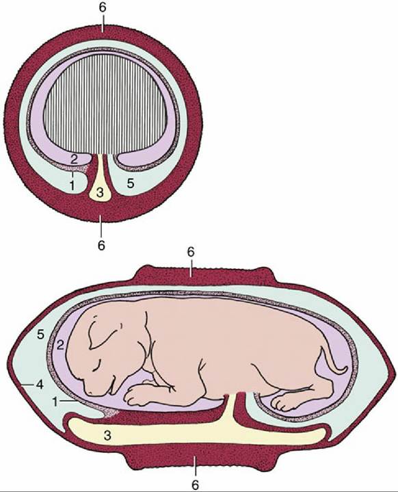
FIG. 15.14 The feline fetal membranes in transverse and longitudinal section, schematic. 1, Amnion; 2, amniotic cavity; 3, yolk sac; 4, chorioallantois; 5, allantoic cavity; 6, zonary placenta.
Initially the uterus enlarges locally, and each conceptus is confined within a globular swelling that is bounded by regions of constriction. The separate ampullae persist until about the 40th day (in a gestation that averages 63 days, measured from the date of ovulation*), when there begins a gradual relaxation of the constrictions, eventually creating an almost uniformly expanded uterus. The positions of the individual fetuses are still obvious on inspection of the exposed organ because the whole thickness of the uterine wall is very vascular at the placental sites. The uterine horns are relatively fixed at their extremities, and when they lengthen, they are forced into loops that first bend cranially from the ovarian attachment before sweeping ventrally, then caudally, to join the body (Fig. 15.15). The pattern of coiling is even more complicated when the litter is large, and radiographs obtained in late pregnancy (when there is mineralization of the fetal skeletons) sometimes show the puppies arranged in a confusing jumble (Fig. 15.16B).
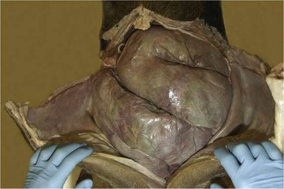
FIG. 15.15 Pregnant uterus of bitch, dominating the abdominal topography.
Pregnancy can be diagnosed through abdominal palpation of round swellings that are approximately 1 cm in diameter from 18 to 21 days of gestation and 2.5 to 4 cm in diameter between 24 and 32 days. From 35 to 45 days of gestation, the swellings enlarge, elongate, and become flaccid and are found ventrally in the abdomen.
For a few days starting from about the 50th day, it is no longer possible to palpate individual swellings, but from the 55th day of gestation individual fetuses are easily palpable.In the later stages of pregnancy abdominal radiographs serve to determine the number of pups in the litter and provide a means of assessing fetal age, thus predicting the date of parturition. Mineralization commences in the axial skeleton by about the 45th day and soon progresses to that of the appendicular skeleton in proximodistal sequence (see Fig. 5.74; Table 15.1). Mineralization of the skeleton of kittens follows the same pattern, but each element makes its appearance a few days earlier than in pups.
Ultrasonography provides an alternative or additional means of diagnosing pregnancy and predicting term. Claims have been made for its success in recognizing uterine enlargement at a very early stage, but confident diagnosis requires a longer wait (perhaps 28 days). Even then, exact litter size cannot be determined. In cats, a gestational sac is visible about days 11 to 14, and fetal cardiac activity is present at day 14.
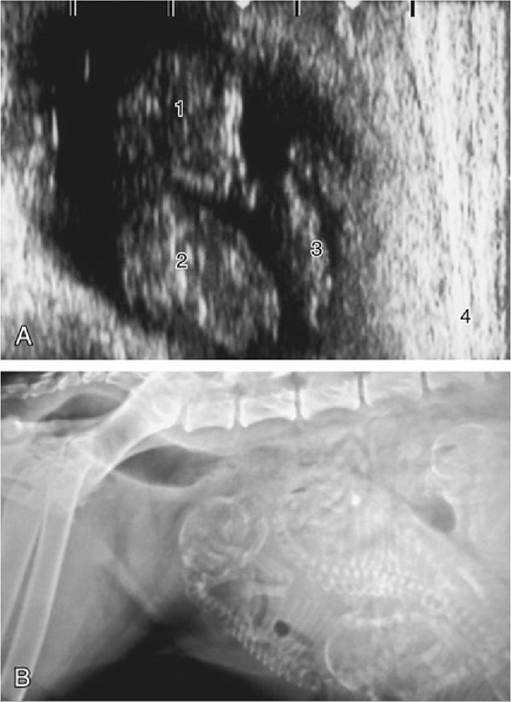
FIG. 15.16 (A) Ultrasonographic (transabdominal) view of a 33-day (after a single mating) Beagle fetus in its ampulla; the scale at top is in centimeters. 1, Head of fetus; 2, thorax of fetus; 3, yolk sac; 4, uterine wall. (B) Radiograph (lateral view) of pregnant bitch with several almost full-term fetuses. Note the gas in the rectum.
» TABLE 15.1
Guide to the Mineralization of Dog Fetuses
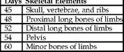
Modified from Concannon P, Rendano V: Radiographic diagnosis of canine pregnancy: onset of fetal skeletal radiopacity in relation to times of breeding, preovulatory luteinizing hormone release, and parturition, Am J Vet Res 44:1506-1512, 1983; and Yaeger AE, Mohammed HO, Meyers-Wallen V, et al: Ultrasonographic appearance of the uterus, placenta, fetus and fetal membranes throughout accurately timed pregnancy in beagles, Am J Vet Res 53:324-329, 1992.
Parturition is facilitated by pelvic rotation at the sacroiliac joints and by elevation of the tail to significantly increase the dimensions of the pelvis. In both dogs and cats some 60% to 80% of fetuses present the head toward the cervix at term. Fetuses tend to be delivered from each horn in alternation, and when each is delivered, the emptied segment of the uterus contracts and brings those littermates left behind closer to the exit. When expelled, each fetus is still attached to its placenta, from which it is freed by the dam's biting through the umbilical cord. The "afterbirth," with which considerable maternal tissue is shed, is normally consumed.
Although less often useful to the clinician, some information on the development of certain external features of fetuses can be found in Tables 15.2 and 15.3.
The cat is sexually mature at 6 to 9 months of age. The proestrus stage, the nonacceptance of a male, lasts 12 to 48 hours. In cats, pea-sized swellings can be palpated at 21 days of gestation. By 28 days, the swellings are firm and are about 2.0 to 2.5 cm in diameter. The uterus is evenly distended during days 35 and 50 and may be difficult to differentiate from pyometra.
Potentially embarrassing mistakes in the determination of the sex of newborn kittens are relatively easily made. The difficulty arises from the orientation of the penis. The orientation brings the anal and genital openings relatively close together in the tom, and the spacing is inconveniently similar to that in the female.