The Vagina, Vestibule, and Vulva (see also pp. 187-189)
The vagina of the bitch is very long (about 12 cm) and extends horizontally through the pelvis before dipping beyond the ischial arch to join the vestibule (see Fig. 5.35/5 and 9).
Apart from the prominent dorsomedian fold that continues the cervix for a short distance, the interior of the undistended organ is obstructed by the irregular folds that end at the junction with the vestibule (Figs. 15.12 and 15.13). The vestibule continues the downward slope of the vagina, requiring that a vaginal speculum or other instrument be passed in a craniodorsal direction to clear the ischial arch before it can be advanced horizontally (see Fig. 5.2). During such examinations the dorsal fold combines with the lateral and ventral vaginal walls to simulate a cervix (pseudocervix).The cranial part of the vestibular floor (of the bitch) displays the tubercle and flanking depressions associated with the opening of the urethra, and the caudal part presents the fossa into which the glans of the clitoris projects (Fig. 15.12/9 and 9'). The functional significance of the urethral tubercle is not known. Darker patches of the lateral walls betray the positions of the vestibular bulbs in the bitch, but the cat has slighter and more diffuse (even insignificant) version. Vestibular glands are present only in the cat.
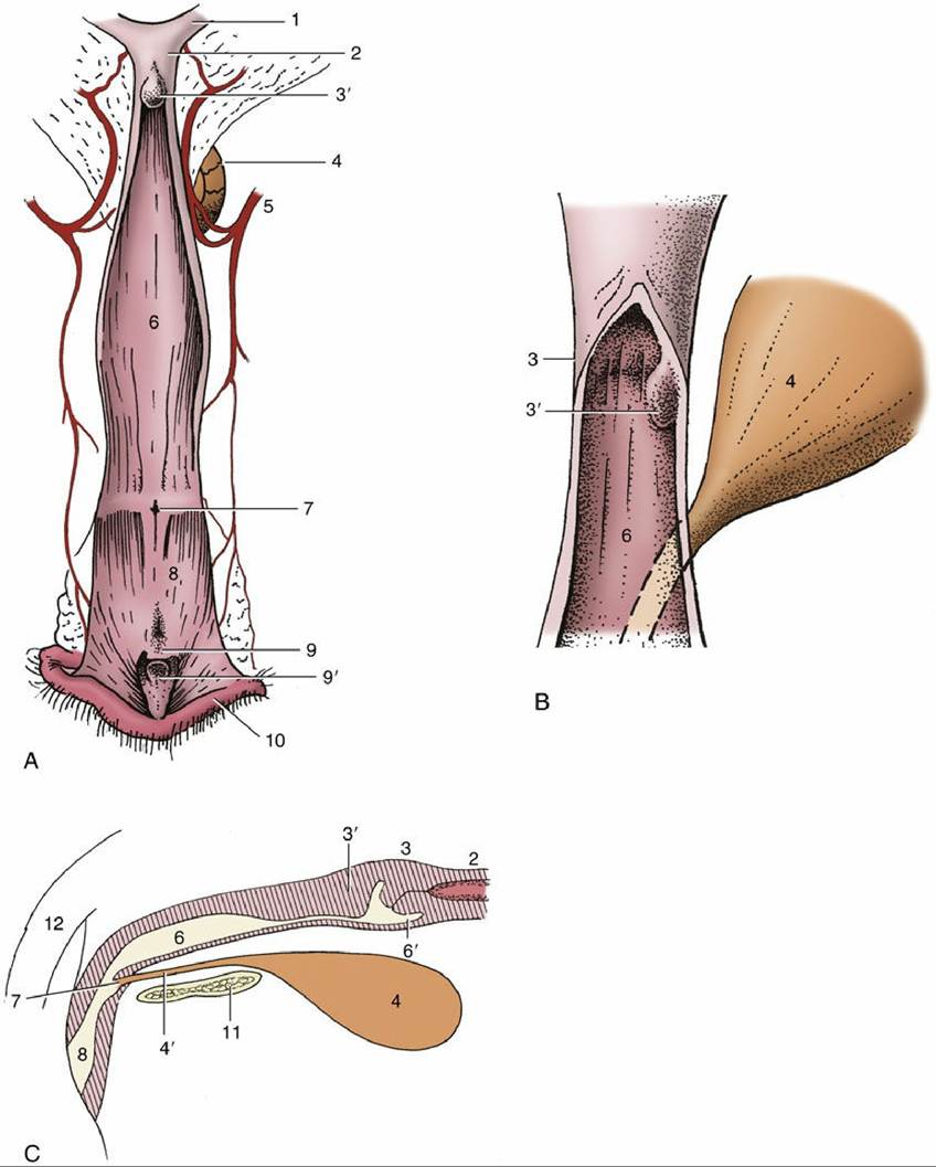
FIG. 15.12 (A) Canine vagina, vestibule, and vulva, opened dorsally. (B) Enlarged view of the cervix. (C) Schematic median section of the organs shown in (A). 1, Right uterine horn; 2, body of uterus; 3, cervix;
3', dorsal fold, which may extend a considerable distance into the vagina; 4, bladder; 4', urethra; 5, vaginal artery; 6, vagina; 6', fornix; 7, external urethral orifice; 8, vestibule; 9, clitoris; 9', clitoral fossa; 10, right labium of vulva; 11, pelvic symphysis; 12, tail.
The thick labia of the vulva meet in a dorsally rounded and ventrally pointed commissure. More lateral folds that are sometimes apparent are believed to be homologous with the labia majora of human anatomy. The crura and body of the clitoris possess a little erectile tissue; the glans is largely of fatty fibrous tissue but sometimes contains a small bone, the os clitoridis. The queen has only a corpus cavernosum clitoridis and not a glans clitoridis.
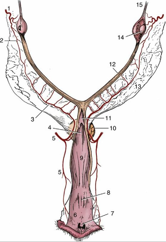
FIG. 15.13 Blood supply of the reproductive organs of the bitch, dorsal view. The right ovarian bursa and the caudal parts of the tract have been opened. 1, Ovarian artery; 2, uterine branch of ovarian artery; 3, uterine artery; 4, dorsomedian fold continuing the cervix; 5, vaginal artery; 6, vestibule; 7, clitoris; 8, external urethral orifice; 9, vagina; 10, bladder; 11, cervix; 12, right uterine horn; 13, broad ligament; 14, right ovary; 15, suspensory ligament of ovary.