Functionally, Pulmonary Blood Vessels Can Be Classified as Alveolar and Extra-Alveolar Vessels
Alveolar vessels are the thin-walled capillaries that perfuse the alveolar septum (Figure 46-1). They are exposed almost directly to the pressure changes that occur in the alveoli during breathing.
Extra-alveolar vessels include the pulmonary arteries, veins, arterioles, and venules. They generally occur together with bronchi in a loose connective tissue sheath called the broncho- vascular bundle. This bundle is bounded by a limiting mem-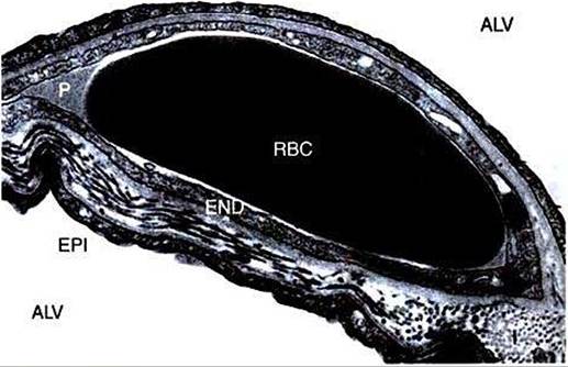
FIGURE 46-1 Transmission electron micrograph of capillary in alveolar septum of horse lung. A red blood cell (RBC) is shown bathed by plasma (P) in a capillary surrounded by endothelium (END). Alveoli (ALV) are on both sides of the septum and separated from the capillary by the epithelium (EPI) and a layer of interstitium (//The interstitium is much thicker on one side of the capillary than on the other. Fluid exchange between the capillary and the interstitium occurs primarily on the thicker side. (Courtesy WSTyIer, Department of Anatomy, University of California-Davis.)
brane to which alveolar septa are attached (Figure 46-2). The behavior of extra-alveolar vessels is determined by pressure changes within the connective tissue space of the broncho- vascular bundle rather than by changes in alveolar pressure.
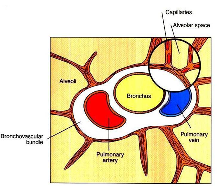
FIGURE 46-2 Diagrammatic representation of the extra-alveolar vessels (pulmonary artery and vein) in the bronchovascular bundle and the alveolar vessels (capillaries) in the alveolar septum.