» General Aspects of Visceral Topography
The greater omentum is extremely well developed and is folded on itself to form a flat sac with superficial and deep leaves that intervene between the intestinal mass and the abdominal floor (see Fig.
3.33). This is the reason that the small intestines are not immediately visible when the abdominal floor is removed (Fig. 14.4). However, the ventral part of the spleen projecting beyond the left costal arch, a part of liver behind the xiphoid process, and the bladder directly before the pubis are visible when the abdominal wall or the floor is removed (Figs. 14.5, 14.6, 14.7, 14.8, and 14.9). The omental bursa exists as a potential space between the leaves. The opening to the omental bursa, the epiploic foramen, is a narrow passage that lies medial to the caudate process of the liver and is bounded dorsally by the caudal vena cava and ventrally by the portal vein.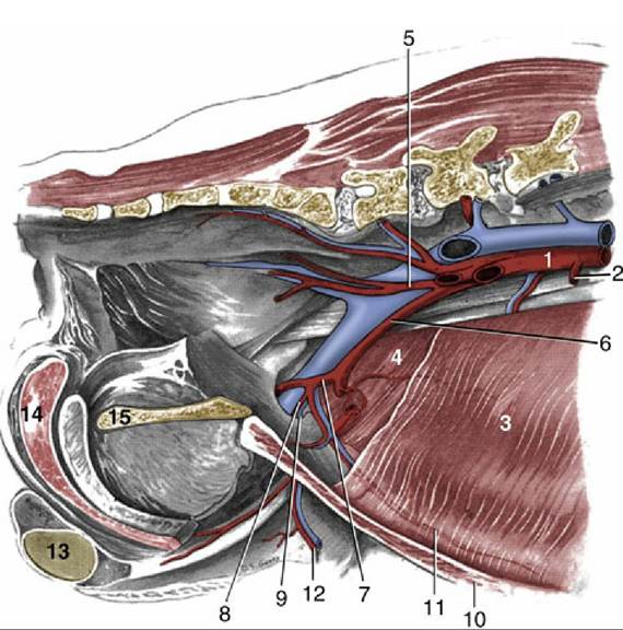
FIG. 14.3 Abdominal wall and pelvic canal of the male dog, showing the breakup of the aorta; medial view. 1, Aorta; 2, caudal mesenteric artery (a.); 3, transversus abdominis; 4, internal abdominal oblique muscle (m.); 5, internal iliac a.; 6, external iliac a.; 7, deep femoral a.; 8, pudendoepigastric trunk; 9, deep inguinal ring; 10, rectus abdominis m.; 11, caudal epigastric a.; 12, external pudendal a.; 13, left testis; 14, bulb of the penis; 15, pelvic symphysis.
Being the dorsal mesogastrium, the greater omentum attaches to the greater curvature of the embryonic stomach, as in other species. It arises from the roof of the abdominal cavity, near the caudal part of the liver and the celiac artery. Close to this attachment the left lobe of the pancreas is enclosed in the omentum. The dorsal attachment of the omentum runs between the esophageal hiatus and the epiploic foramen.
At this point the greater omentum continues as mesoduodenum, in which the right lobe of the pancreas is situated. The omental bursa is connected to a hilus of the spleen by the gastrosplenic ligament. The omentum is connected to the descending colon caudally by the omental veil.The superficial leaf of greater omentum (see Fig. 3.33/14) passes caudally from its attachment, in direct contact with the ventral abdominal wall to reach the bladder, where it is reflected dorsally to become the deep leaf (see Fig. 3.33/13). This structure runs forward between the superficial leaf and coils of the jejunum; at the cranial end of the jejunum it passes dorsally, against the caudal (visceral) surface of the stomach, to reach the left lobe of the pancreas, which it encloses and by means of which it gains the roof of the abdominal cavity. The right border of the omental sac is ventral to the descending duodenum. The left extends more dorsally to the level of the kidney and sublumbar
muscles and is complicated by an attachment to a hilus of the spleen. The part of the omentum extending between the left crus of the diaphragm and the splenic hilus may be known as the phrenicosplenic ligament. The more generous part between the stomach and hilus forms the gastrosplenic ligament. As a further complication, a sagittal fold (omental veil) with a caudal free border connects the deep leaf with the left surface of the descending mesocolon. The greater omentum always contains fat., which is first deposited along the small omental vessels, giving the structure a lacy appearance; however, in obese dogs (less so in cats) it forms a more or less continuous layer.
FIG. 14.4
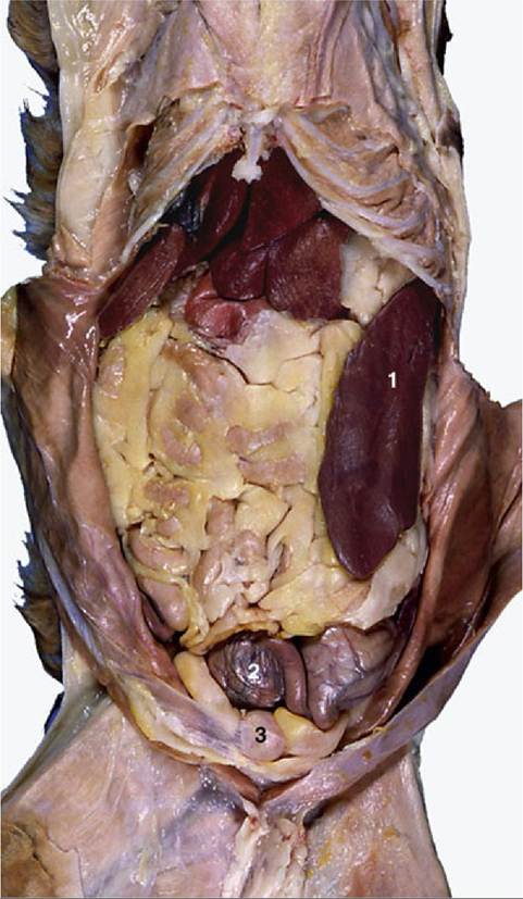
Ventral view of feline abdominal viscera; intestinal loops are concealed by fat-filled greater
omentum. 1, Spleen; 2, part of gravid uterus with two ampullae; 3, bladder.
The lesser omentum is considerably wider than the short space it has to bridge between the lesser curvature of the stomach and the liver. It blends on the right with the mesoduodenum, the bile duct marking the boundary between the two. The papillary process of the liver is loosely enveloped by the lesser omentum. The portion of the lesser omentum between the liver and the duodenum is also called the hepatoduodenal ligament, and the portion between the liver and the stomach is called the hepatogastric ligament.
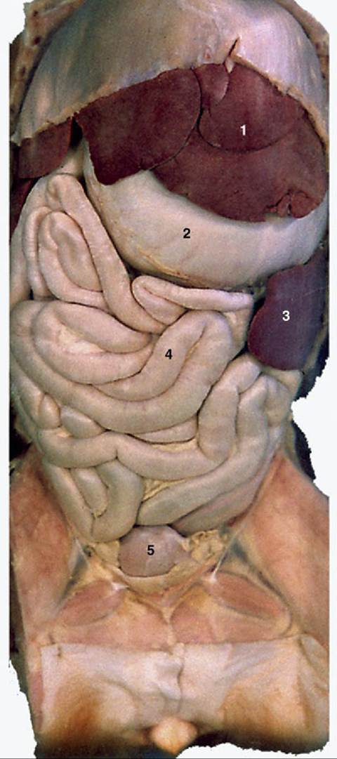
FIG. 14.5 Abdominal viscera of the dog after removal of the greater omentum. 1, Liver; 2, stomach; 3, spleen; 4, small intestine; 5, bladder.