» The Spleen
The spleen (see also p. 249) in dogs and cats is an elongated, roughly dumbbell-shaped organ that lies more or less vertically against the left abdominal wall (Fig. 14.10A/4). Its position is much influenced by the distention of the stomach (and by its own capacity to become engorged).
The dorsal end reaches the left crus of the diaphragm, passing between the gastric fundus and the cranial pole of the left kidney under cover of (usually) the last two ribs. The larger ventral end may cross the ventral midline, to reach under the costal cartilages of the right side. It then provides a dense triangular shadow on the abdominal floor in lateral radiographs (Fig. 14.11A/3). A similar shadow between the stomach and left kidney may reveal the position of the organ in ventrodorsal radiographs. In the cat, the ventral part of the spleen is always located outside the rib cage. The parietal surface makes contact (in dorsoventral sequence) with the diaphragm, costal arch, and abdominal muscles. The visceral surface is divided by a hilar ridge into a cranial strip related to the stomach and a caudal strip related to the left kidney and intestine.FIG. 14.6
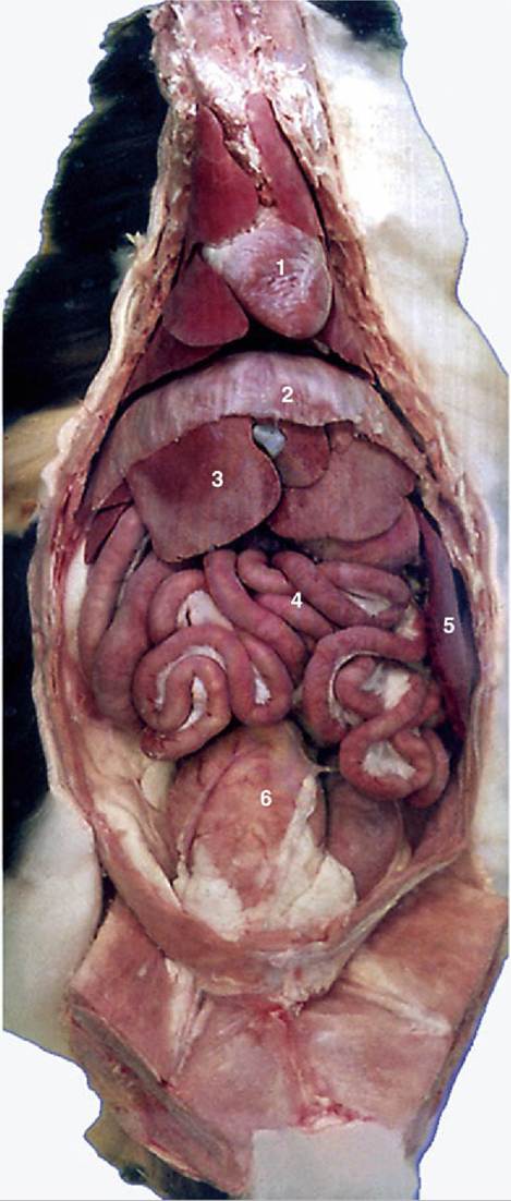
Ventral view of feline viscera after removal of omentum. 1, Heart; 2, diaphragm; 3, liver; 4, intestine; 5, spleen; 6, bladder.
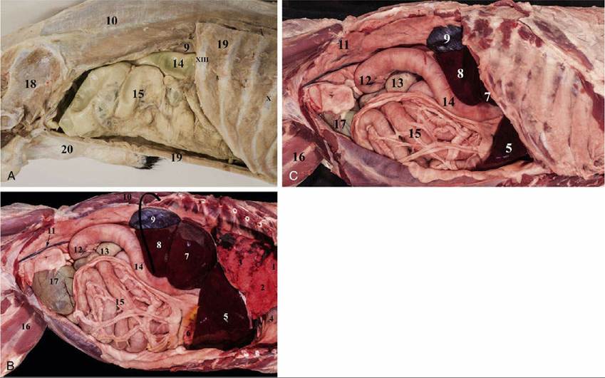
FIG. 14.7 Abdominal cavity of the dog with right abdominal wall removed (A and B) and the ribs removed (B). The black line in B indicates the position of the last rib. 1. Cranial lobe of the right lung; 2. Middle lobe of the right lung; 3. Caudal lobe of the right lung; 4. Heart; 5. Right medial lobe of the liver; 6. Stomach, greater curvature; 7. Right lateral lobe of the liver; 8.
Caudate process of the caudate lobe of the liver; 9. Right kidney; 10. Longissimus lumborum muscle; 11. Testicular artery and vein in the genital fold; 12.Caudal duodenal flexure; 13. Cecum; 14. Descending duodenum; 15. Jejunal loops, covered by the greater omentum; 16. Sartorius muscle (cranial and caudal part); 17. Urinary blader; 18. Ilium (pelvis); 19. Abdominal muscles (cut); 20. Bulbus glandisX, XIII: ribs
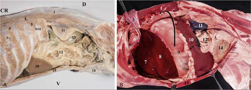
FIG. 14.8 Abdominal cavity of the dog viewed from left side (A) and with ribs removed (B): 1. Longissimus lumborum muscle; 2. Sympathetic trunk; 3. Diaphragm (reflected); 4. Iliocostalis thoracis muscle; 5. Serratus dorsalis muscle (caudal part); 6. Esophagus and the dorsal trunk of the vagus nerve; 7. Left medial lobe of the liver; 8. Left lateral lobe of the liver; 9. Stomach; 10. Spleen; 11. Left kidney; 12. Jejunal loops, covered by the greater omentum; 13. Descending colon 14. Urinary blader; 15. Abdominal muscles (cut);black line represents the last (XIII.) rib
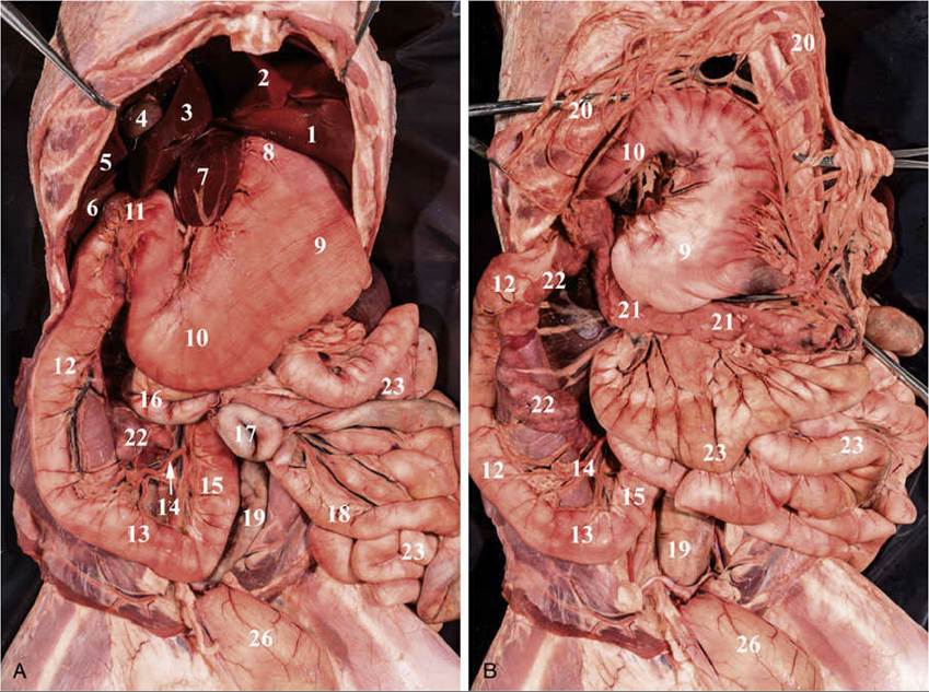
FIG. 14.9 Abdominal cavity of the dog, jejunum flipped to the left (A), stomach and the greater omentum
flipped cranially (B), and small intestine removed, colon flipped to the right (fresh specimens): 1. Left
lateral lobe of the liver; 2. Left medial lobe of the liver; 3. Quadrate lobe of the liver; 4. Gallblader; 5. Right medial lobe of the liver; 6. Right lateral lobe of the liver 7. Papillary process of the caudal lobe of the liver
(covered by the lesser omentum); 8. Stomach, cardia; 9. Stomach, fundus 10. Stomach, pyloric part; 11. Cranial duodenal flexure; 12. Descending duodenum; 13. Caudal duodenal flexure; 14. Caudal
pancreaticoduodenal artery and vein; 15. Ascending duodenum ; 16. Ascending colon; 17. Cecum; 18. Ileum; 19. Descending colon; 22. Pancreas, right lobe; 23.
Jejunal loops, flipped to the left side; 26.Urinary blader.
The wide gastrosplenic ligament attaches the spleen to the greater curvature of the stomach, affecting the latter's mobility and location. When the stomach enlarges, the spleen is displaced caudally and ventrally, reaching the pelvic inlet. It may then be palpated through the abdominal wall.
The blood vessels also exert another restraining influence on the spleen. The splenic artery and vein pass (as several divergent branches) to the dorsal end of the spleen. The splenic artery arises as a branch of the celiac artery, and before reaching the spleen, it gives off branches to the left limb of the pancreas. The left gastroepiploic vessels are detached about the middle of the hilus and cross to the greater curvature of the stomach within the gastrosplenic ligament (Fig. 14.12/3 and 11). The splenic lymph nodes lie near the splenic vessels, a few centimeters distant from the organ. The spleen has efferent lymphatic vessels (which follow large arteries) but no afferent vessels.
Spleen Function: The spleen serves as an important blood reservoir in the dog and cat, and its size and weight therefore vary widely (Fig. 14.6). The spleen in a resting dog or cat contracts and relaxes rhythmically because of the presence of many smooth muscle fibers throughout the organ. These fibers relax when anesthetics are used, resulting in marked splenic enlargement, and contract because of stress or injection of catecholamines, expelling free blood cells and plasma from the red pulp. The spleen has no parasympathetic nerve supply.
Rupture of the spleen is not uncommon after traffic accidents, but fortunately the organ may be removed without risk to life. The relatively loose attachment of the spleen to the stomach facilitates access to the vascular supply at surgery (splenectomy). During splenectomy, it is important to spare branches of the splenic artery that contribute to the left gastroepiploic artery, which is essential to the integrity of the greater curvature of the stomach (see Figs. 3.39 and 14.12).
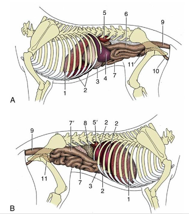
FIG. 14.10 Visceral projections on the (A) left and (B) right canine abdominal walls. 1, Diaphragm; 2, liver; 3, stomach; 4, spleen; 5 and 5', left and right kidneys; 6, descending colon; 7, small intestine; 7', descending duodenum; 8, pancreas; 9, rectum; 10, female urogenital tract; 11, bladder.