General Considerations
The stomach is composed of four chambers—rumen, reticulum, omasum, and abomasum—through which the food passes successively (Fig. 28.7). The first three, collectively known as the forestomach (proventriculus), are developed to cope with the complex carbohydrates that form so large a part of the normal diet of ruminants, and only the last chamber is comparable in structure and function to the simple stomach of most other species.
All are derived, however, from the gastric spindle of the embryo (Fig. 28.8).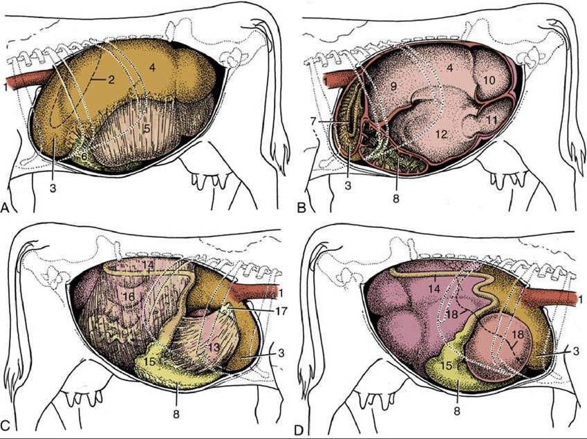
FIG. 28.4 Topography of the abdominal viscera. (A) Relationship of abdominal viscera to the left abdominal wall. (B) The interior of the stomach seen from the left. (C) Relationship of abdominal viscera to the right abdominal wall; the liver has been removed. (D) Position of the parts of the stomach seen from the right. 1, Esophagus; 2, outline of spleen; 3, reticulum; 4, dorsal sac of rumen; 5, ventral sac of rumen, covered by superficial wall of greater omentum; 6, fundus of abomasum, covered by superficial wall of greater omentum; 7, reticular groove; 8, body of abomasum; 9, atrium ruminis; 10, caudodorsal blind sac; 11, caudoventral blind sac; 12, ventral sac of rumen (opened); 13, omasum, covered by lesser omentum;
14, descending duodenum; 15, pyloric part of abomasum; 16, greater omentum covering the intestinal mass; 17, lesser omentum cut away from the liver; 18, position of caudoventral border of liver.
The topography of the ruminant abdomen is dominated by the enormous development of the stomach, which in adult cattle almost fills the left half of the cavity and occupies a substantial portion of the right (Figs. 28.9, 28.10, 28.11, and 28.12 and see Fig. 27.1). Its capacity measures about 60 L. This amount, which is much more modest than many estimates, may be apportioned between the various chambers as follows: rumen, 80%; reticulum, 5%; omasum, 8%; and abomasum, 7%.
The proportions in small ruminants are somewhat different, being perhaps 75% rumen, 8% reticulum, 4% omasum, and 13% abomasum. The relative volumes are fairly constant in the short term because the enormous storage capacity of the first chambers and the more or less continuous passage of ingesta into the distal parts minimize the effects of intermittent feeding.The different chambers are identifiable as expansions of the foregut spindle in the early embryo.
They increase at unequal rates throughout the embryonic and fetal periods as first one takes the lead and then another. At one stage the fetal stomach has an almost adult configuration, but during the last months of intrauterine life the abomasum outstrips the others; at birth it accounts for more than half the weight and capacity of the entire organ—which is appropriate because it is the only part that has an immediate function to perform. The postnatal changes through which the adult proportions and topography are acquired are described later (p. 680).
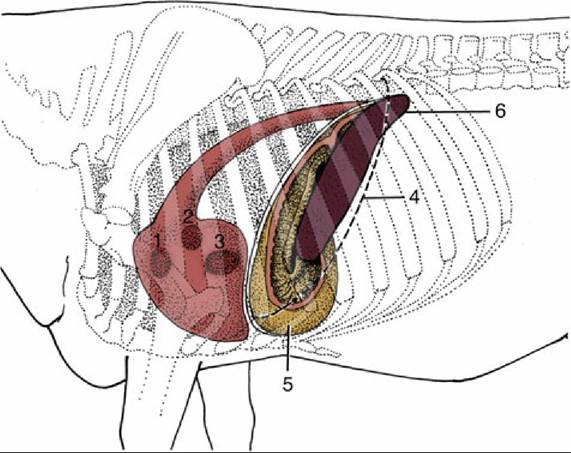
FIG. 28.5 Left lateral projection of certain organs on the bovine thoracic wall. 1, Pulmonary valve; 2, aortic valve; 3, left atrioventricular valve; 4, position of basal border of the lung; 5, reticulum, opened (note position of reticular groove); 6, spleen.
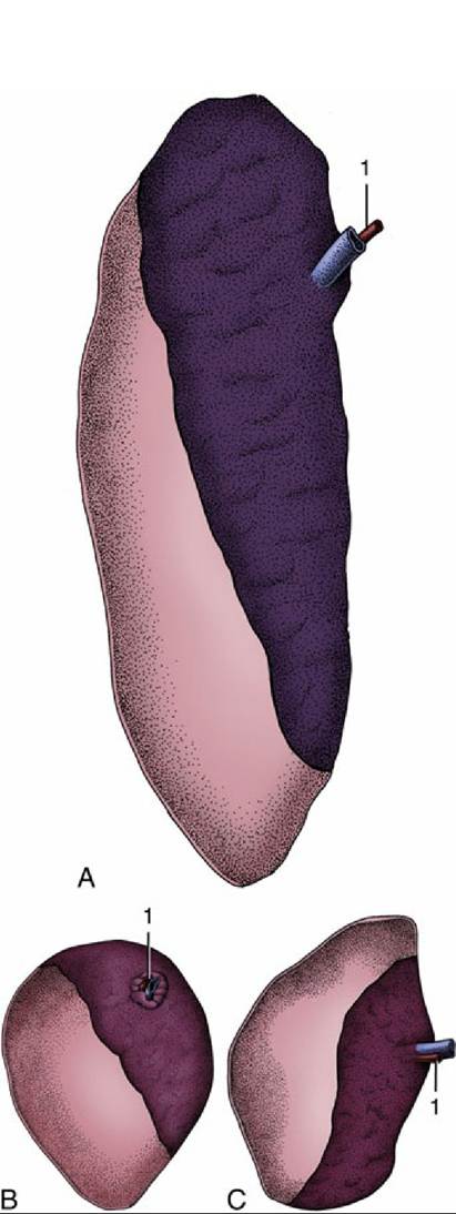
FIG. 28.6 The spleens of (A) cattle, (B) sheep, and (C) goats; visceral surface. The Craniodorsal area is bare. 1, Splenic artery.
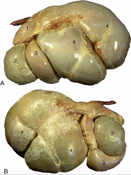
FIG. 28.7 (A) Bovine stomach, left side. (B) Bovine stomach, right side. 1, Reticulum; 2, omasum; 3, abomasum; 4, rumen.
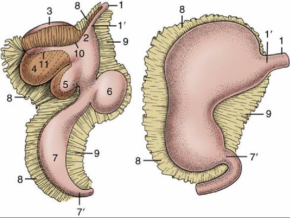
FIG. 28.8 The attachments of the greater and lesser omenta on the developing ruminant stomach. The simple stomach to the right shows the correspondence of its parts to the compartments of the ruminant stomach. 1, Esophagus; 1', cardia; 2, atrium ruminis; 3, dorsal sac of rumen; 4, ventral sac of rumen; 5, reticulum; 6, omasum; 7, abomasum; 7', pylorus; 8, greater omentum; 9, lesser omentum; 10, part of greater curvature corresponding to the right longitudinal groove of the rumen; 11, part of greater curvature corresponding to the left longitudinal groove of the rumen.
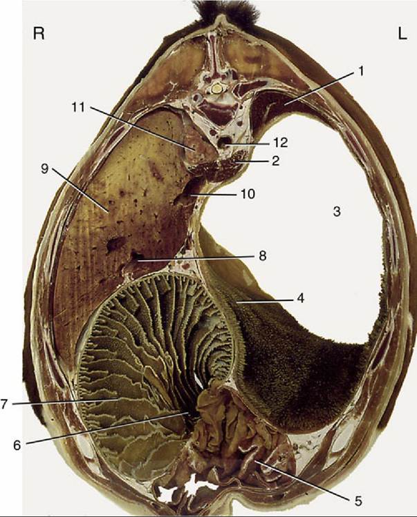
FIG. 28.9 Transverse section of the bovine trunk at the level of the 10th thoracic vertebra. 1, Spleen; 2, crura of diaphragm; 3, atrium ruminis; 4, cranial pillar; 5, abomasum; 6, omasoabomasal opening; 7, omasum; 8, portal vein; 9, liver; 10, caudal vena cava; 11, right lung; 12, aorta; L, left side; R, right side.