» The Spleen
A general impression of the visceral topography should be obtained from Fig. 28.4 before the individual organs are considered.
The flat oblong spleen of adult cattle is about 45 cm long and 12 cm wide.
It is situated over the craniodorsal part of the rumen, against the left half of the diaphragm, and is attached to both these organs by the gastrosplenic ligament and the phrenicosplenic ligament, respectively. Its upper end lies under the dorsal ends of the last few ribs, and its axis extends ventrally, with a slight cranial inclination, across the line of the ribs to end in the region of the seventh costochondral joint (Figs. 28.4A/2 and 28.5/6). In most animals the lower end passes onto the reticulum, which brings risk of involvement in the common abscesses and perforations of that organ. The upper part of the spleen is retroperitoneal: the line of serosal reflection runs cranioventrally over both parietal and visceral surfaces. The hilus is confined to the dorsocranial angle of the medial side, and to reach this site, the splenic vessels must first pass over the roof of the rumen.The capsule contains little muscle, and physiologic variation in spleen size is therefore rather restricted. Occasionally an enlarged spleen may extend behind the last rib in the angle between this and the lumbar spine, but for practical purposes the spleen may be regarded as out of reach for palpation or percussion. Access for a biopsy is normally made through the upper end of the 11th intercostal space and involves little risk of injury to the lung, particularly if the needle is introduced during expiration.
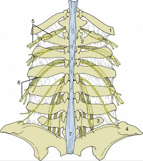
FIG. 28.3 Relationship of the lumbar spinal nerves to the transverse processes of the bovine lumbar vertebrae. 1, Last rib; 2, first lumbar vertebra; 3, sixth lumbar vertebra; 4, coxal tuber; 5, dorsal and ventral branches of the 13th thoracic nerve (the ventral branch is partly stippled); 6, dorsal and ventral branches of second lumbar nerve; 7, supraspinous ligament.
The spleen has a relatively soft consistency. Its color varies considerably, tending to be steel blue in cows and more reddish in males and younger animals. The division of the pulp into red and white areas is very obvious. The white corpuscles are somewhat larger than pinheads.