General Visceral Efferent Neurons
These motor pathways (GVE) belong to the autonomic system and are divided into the sympathetic and parasympathetic divisions. The sympathetic system is responsible for widespread visceral responses required in emergency, the so-called ‘fight or flight response.
The parasympathetic system is responsible for the visceral responses that are required for the maintenance of the body's reserves and tends to act locally. The two systems often act antagonistically, although some organs are innervated by the sympathetic system only (e.g. the adrenal medulla, sweat glands and many blood vessels).The two systems act through different transmitter substances at their effector endings; these are noradrenaline or adrenaline at sympathetic effector endings and acetylcholine at parasympathetic endings.
11.4.1 Sympathetic motor pathways (Figures 11.1, 11.2, 11.3, 11.4 and 11.7)
The sympathetic outflow from the central nervous system (CNS) is limited to the thoracic and rostral lumbar segments of the spinal cord; it extends to as far as L1 to L5 depending on species. The cell bodies of these preganglionic sympathetic neurons are located in the lateral horn of the spinal cord and represent the entire outflow of sympathetic motor pathways to the whole of the body. Their myelinated axons leave the cord by the ventral root, enter a white ramus communicans and end in one of a chain of vertebral ganglia (ganglia of the sympathetic trunk, NAV).
The preganglionic neurons form synapses with several postganglionic neurons in the vertebral ganglia. In fact the ratio of pre- to postganglionic neurons is high, about 1:20 or more. The axons of the postganglionic neurons return to the spinal nerves via an unmyelinated grey ramus communicans.
In the abdomen (originating in segments T6-T10 in the cat) the axons of the preganglionic neurons pass straight through the vertebral ganglia and contribute to the great splanchnic nerve.
The axon ends by entering a prevertebral ganglion,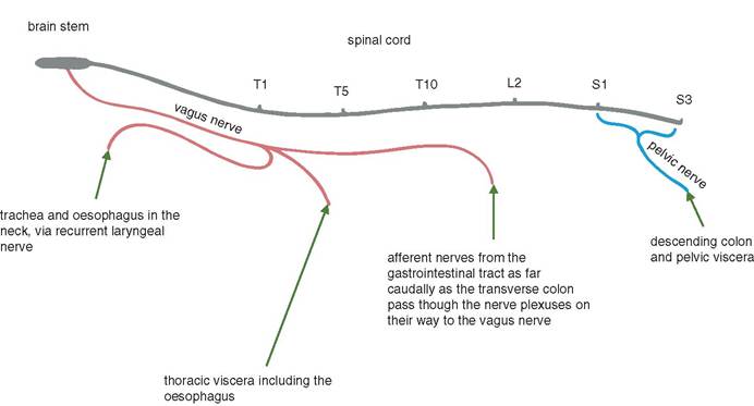
Figure 11.1 Summary of the afferent pathways from the viscera, excluding pain. The non-pain sensory nerve pathways from the thoracic, abdominal and pelvic viscera travel to the neuraxis in the vagus and pelvic nerves.
T6
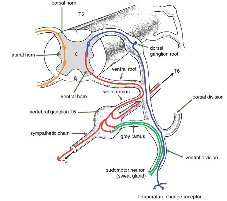
Figure 11.2 Diagram to show the sympathetic motor pathway of the somatic region of the abdomen.
The example shows a receptor sensitive to temperature change, and the effector is a sudomotor
neuron.
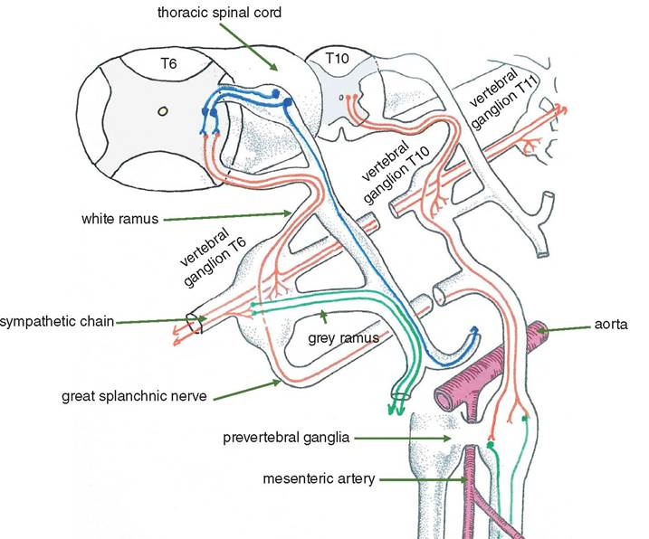
Figure 11.3 Diagram of the sympathetic motor pathway to the viscera. Refer to Figure 11.2 for the colour code. The preganglionic nerves originating in segments T6-T10 (in the cat) pass through the vertebral ganglia and enter the greater splanchnic nerve. They continue into the coeliacomesenteric plexus to synapse in a prevertebral ganglion with many postganglionic neurons, which are then distributed to target organs.
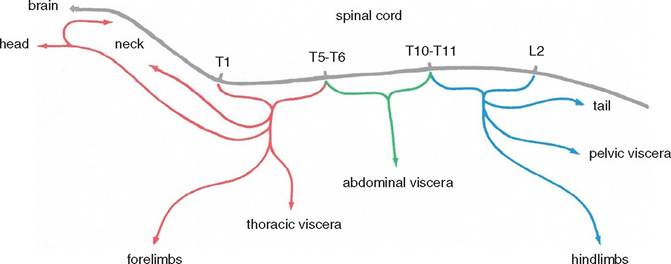
Figure 11.4 Summary of the motor outflow of the sympathetic system. The diagram is based on the cat, in which a total of 15 segments (T1 to L2 inclusive) contribute to the sympathetic outflow from the neuraxis. These 15 segments can be divided functionally into three groups, each consisting of five segments. These three groups supply particular regions of the body, essentially as shown.
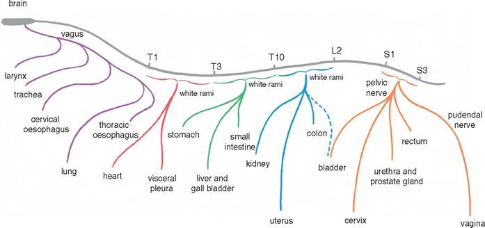
Figure 11.5 Summary of the pain pathways from the viscera. The great majority of the pain pathways from the viscera are projected to the CNS by axons that travel in sympathetic nerves. Topographically these pain pathways tend to resemble the motor pathways of the sympathetic system (see
Figure 11.4).
Thus, both the motor pathways and the pain pathways of the thoracic viscera travel mainly in the sympathetic nerves relating to the segments T1—T5; likewise both the motor pathways and the pain pathways to the abdominal viscera travel mainly in the sympathetic nerves relating to the segments T6-T10. However, the pain pathways from the pelvic viscera break with this principle by projecting to the CNS via the pelvic and pudendal nerves rather than via the sympathetic nerves relating to segments T11-L2. The pain pathways from the respiratory tract including the lung form another exception by travelling in the vagus.where it forms numerous branches that synapse with about 20 postganglionic neurons. Again the ratio of pre- to postganglionic neurons is therefore about 1:20, as in the vertebral ganglia.
The prevertebral ganglia (ganglia of the autonomic plexuses, NAV) arise as paired primordia that fuse to varying extents. They lie on the ventral aspect of the dorsal aorta, and the left ganglion tends to fuse with that on the right.
There are a number of prevertebral ganglia in the abdomen. The most important are the coeliac, cranial mesenteric and caudal mesenteric ganglia. The coeliac and cranial mesenteric ganglia become fused in many species to form a composite coeliacomesen- teric ganglion that supplies postganglionic nerves to the abdominal viscera. These sympathetic nerves travel to their target organs along the arteries, which supply these organs, e.g. the axons supplying the wall of the stomach travel along the branches of the coeliac artery. The caudal mesenteric ganglion distributes sympathetic postganglionic axons via the hypogastric nerves to the pelvic viscera, e.g. the urogenital organs, rectum, etc.
11.4.2 The prevertebral ganglia (Figures 11.1 and 11.2)
The prevertebral ganglia contain postganglionic neurons of three functional types:
• Vasomotor to blood vessels of the gastrointestinal tract
• Motor to the smooth muscle in the wall of the gastrointestinal tract
• Motor to the glands of the of the gastrointestinal tract.
The vasomotor fibres are very numerous and strongly influence the systemic arterial blood pressure by constituting a variable resistance to the output of the left ventricle. For example, if the blood pressure falls because of haemorrhage, a reflex vasoconstriction of the splanchnic circulation occurs.
The sympathetic innervation of the smooth muscle of the gut causes the sphincters to close and the intestine wall to relax. Hence the movement of ingesta is arrested.
Also, in line with a ‘fight or flight' response, the sympathetic innervation of the glands of the intestine promotes inhibition.
11.4.3 Sympathetic transmitter substances
Preganglionic sympathetic axons release acetylcholine at their endings and are called cholinergic fibres. Many interneurons in the CNS also release acetylcholine at their endings.
Most postganglionic sympathetic axons release adrenaline or noradrenaline at their endings; they are called adrenergic fibres. There are some exceptions, e.g. the sympathetic postganglionic nerves that innervate merocrine sweat glands and sympathetic vasodilator fibres are cholinergic.
The endocrine cells of the adrenal medulla are derived embryologically from neural crest tissue and release noradrenaline or adrenaline. The medullary cells are the equivalent of postganglionic cells that are without axons.
11.4.4 Pain pathways from the abdominal viscera (Figure 11.5 and 11.6)
Most of the pathways of visceral pain are carried in the sympathetic nerves. The visceral peritoneum and the gastrointestinal tract are insensitive to crushing and cutting, but they are painful when stretched or subject to muscular spasm. These GVA pathways accompany the sympathetic GVE pathways and pass through the white rami on their way to the dorsal root ganglia of segments T5-L2. However, the pain pathways from the pelvic viscera travel in the pelvic and pudendal nerves via the dorsal root ganglia to the sacral segments of the spinal cord.
11.4.5 Parasympathetic motor pathways (Figure 11.7)
In general the function of the parasympathetic motor pathways is to conserve the reserves of the organism.
Both the pre- and the postganglionic parasympathetic endings release acetylcholine.The outflow of parasympathetic preganglionic pathways is restricted to the sacral and cranial regions of the CNS. However, the vagus nerve provides the pathway for the parasympathetic nerve supply to the gastrointestinal organs. The parasympathetic postganglionic neurons are located between the longitudinal and circular muscle layers of the gut and are called the myenteric, or Auerbach’s plexus. There is also some sympathetic input to this plexus and even a sensory component.
The sacral outflow originates from the sacral segments of the spinal cord and is related to the number of sacral vertebrae in each species, e.g. S1, S2 and S3 in the cat and dog, S1-S5 in the horse and ox. The cell bodies of the preganglionic neurons are located at a site that corresponds to the lateral horn of the spinal cord. The axons are combined as the pelvic nerve (renamed as the splanchnic nerve in NAV). Its main
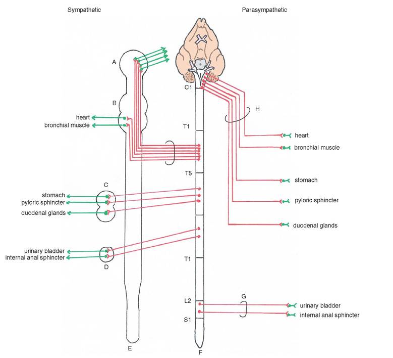
Figure 11.6 Diagram of the sympathetic and parasympathetic motor pathways to the abdominal and pelvic viscera. A = cranial cervical ganglion; B = stellate ganglion; C = Coeliacomesentreic ganglion; D = caudalmesenteric ganglion; E = sympathetic chain; F = spinal cord; G = pelvic nerve; H = vagus nerve; J = great splanchnic nerve.
function is to open smooth muscle sphincters in the pelvic viscera and, at the same time, to contract the muscle in the walls of the bladder and rectum. The pelvic nerve also contains parasympathetic vasodilator nerve fibres to the erectile tissue of the penis.
Both the anus and bladder also have external sphincters of striated muscle that are innervated by GSE fibres. The external anal sphincter is innervated by the pudendal nerve.
11.5