GROWTH AND CYCLICAL CHANGES
The growth of the reproductive organs, isometric in the very young, accelerates in response to the production of ovarian hormones after the initiation of the estrus cycle, generally when a heifer is about 8 to 10 months old.
The cumulative effects of a few cycles produce a striking increase in the dimensions and a clearer differentiation of the component tissues of the tract.In each cycle a follicle becomes identifiable on rectal examination about the 16th day and attains its full size a couple of days later. Its rupture is preceded by a reduction in internal pressure, recognizable on rectal palpation; the clot that succeeds the moderate ensuing hemorrhage is soon replaced by a corpus luteum. This reaches its maximal size, approximately that of the follicle it replaces, after about a week; regression then begins, and by the 21st day, the time of the next estrus, it has already shrunk by about two thirds. It is eventually replaced by a scar. The waxing and waning of the corpus luteum are marked by color changes progressing from brown to ochre and then through orange, brick red, and dirty white in regression.
The ampulla becomes noticeably wider after ovulation, when the sphincter action of the isthmus delays
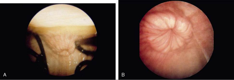
Figure 29-16 The appearance of the vaginal part of the bovine cervix during pregnancy (A) and during estrus (B).
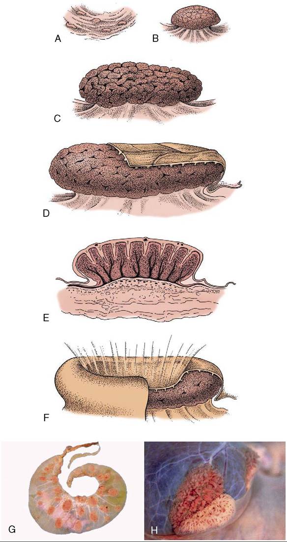
Figure 29-17 A-E, Development of caruncles in the wall of the bovine uterus. A, Caruncle in a nongravid uterus. B, Caruncle in a 2-week gravid uterus. C, Caruncle in a 6-month gravid uterus. D, Caruncle near term, covered in part by a cotyledon (fetal tissue). E, Section of a placentome. F, Placentome of a sheep.
G, Cotyledonary placenta (ruminant). H, Partial separation of maternal and fetal parts of placentome (cow).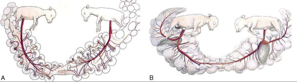
Figure 29-18 A, Twin bovine pregnancy showing separate circulations. B, Twin bovine pregnancy showing conjoined circulations (freemartin development possible).
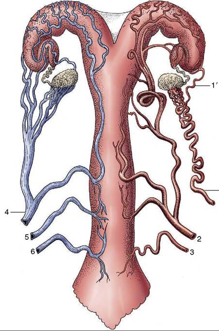
Figure 29-19 Semischematic, ventral view of the blood supply to the bovine reproductive tract (cow). The arteries are depicted on the right side, the veins on the left. 1, Ovarian artery; 1', uterine branch; 2, uterine artery; 3, vaginal artery; 4, ovarian vein; 5, accessory vaginal vein; 6, vaginal vein.
the entry of the egg into the uterus. The uterine changes that commence in proestrus and continue into metestrus involve hyperemia and edema thickening the endometrium; the moderate hemorrhage that sometimes accompanies their subsidence appears to be the origin of the increasing pigmentation of the uterine wall of older animals.
An increase in the size, complexity, and activity of the endometrial glands culminates a week or so after ovulation. The activity of the myometrium, whether spontaneous or in response to external stimuli, is greatest immediately before and during estrus.
The greater activity of the cervical mucosa during estrus spreads to the mucosa that lines the cranial part of the vagina. The transparent mucus of low viscosity that is produced is eventually discharged and may be tinged with blood when bleeding at metestrus is pronounced. There is no distinct cycle of cornification of the vaginal epithelium.
The bovine estrus cycle is repeated at intervals of 21 days. The small ruminants are seasonally polyestrous, largely in the fall and early winter; the cycle lasts 16 or 17 days in sheep and 20 in goats.