GESTATION AND PARTURITION
Gestation lasts 280 days in cattle, 147 days in sheep, and 154 days in goats. During this time every part of the reproductive system shows some changes, but obviously the most striking are in the uterus, which increases its weight 15-fold (100-fold when its content is included).
The ovary is distinguished by the presence of the corpus luteum of pregnancy, which persists beyond the life span of the periodic body of the infertile cycle. Its survival is not always accompanied by total suppression of follicular activity; a few cows come into heat and ovulate in early pregnancy. The corpus luteum is not necessary for the support of pregnancy during the last 3 months and usually begins to regress about a month before term (Figures 29-20 and 29-21).
The progestational changes that are part of every cycle persist and intensify in the presence of an embryo. Although the blastocyst is initially confined to one horn, the membranes soon spread to the other; however, the embryo, later the fetus, is almost invariably restricted
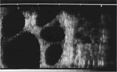
Figure 29-20 Ultrasonographic transrectal scan of the ovary of a cow that was stimulated with gonadotropin to induce superovulation. The black spots represent sections of large tertiary follicles.
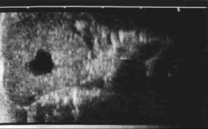
Figure 29-21 Ultrasonographic transrectal scan of a corpus luteum of a cycling cow; the corpus is marked by a cavity (black spot).
unilaterally, and a developing asymmetry is one of the first detectable signs of pregnancy. The amniotic sac becomes palpable about the 30th day, the fetus itself about the 70th. The caruncles of the gravid horn gradually increase from low, smooth-surfaced bumps to become large, pedunculated swellings with surfaces pitted for the reception of the chorionic villi; by term the largest may attain the size of a clenched fist (Figures 29-17, 29-22, and 29-23).
Those in the nongravid horn later also enlarge but to a lesser degree.The enlargement of the uterus does not affect all parts equally. The lesser curvature, being tethered by the broad ligament, most resists expansion, which causes the horn to alter shape: the greater curvature and adjacent parts grow away from the attachment. Hypertrophy of the tissues of the broad ligament restrains the uterus from sinking into the abdomen for a time, but by the third month this resistance is overcome and the uterus begins to slip forward over the abdominal floor. The supply of blood to the gravid uterus is necessarily greatly increased; all uterine vessels contribute to this,
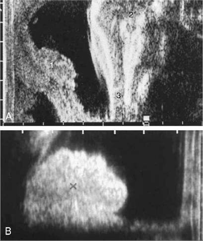
Figure 29-22 A, Transrectal ultrasonic view of a placen- tome (1) and fetal head (2) at 3 months' gestation. The two lower jaws of the fetus are at 3. B, Transrectal ultrasonic view of a placentome (+) at 5 months' gestation. Ultrasonic views of placentomes are diagnostic of pregnancy if the fetus itself cannot be visualized.
but the major role is played by the uterine artery of the gravid side, which increases in diameter from a few millimeters to a centimeter or more. It loses its flexuous character and now passes forward into the abdomen, where it is easily found on palpation against the ilium; identification is assisted by the characteristic vibration (fremitus) it now displays.
The topography is not the same in every pregnancy. The enlarging uterus usually enters the supraomental recess but sometimes may slip forward against the right or the left flank. As it expands, it sinks within the abdomen and for a time passes out of reach of a hand within the colon; this inability to reach the uterus at about the fifth month is as diagnostic of pregnancy as its palpable enlargement at earlier and later times. The descent into the abdomen stretches the vagina and carries the cervix over the pubic brim. Toward term, the uterus occupies most of the ventral and right sections (in the common arrangement) of the abdomen, which raises the rumen dorsally and crushes the intestines upward (Figure 29-24). It makes contact with the liver and diaphragm, on which it exerts increasing pressure.
During the first months the calf enjoys freedom to move and adjust position within the amniotic fluid, but as pregnancy continues, it is forced to adapt to the form and dimensions of the uterine horn.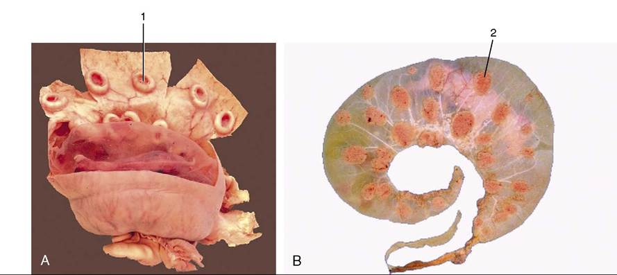
Figure 29-23 A, A gravid uterus, partly opened. B, A bovine fetus within its membranes. The villi are mainly restricted to the cotyledons. 1, Caruncle; 2, cotyledon.
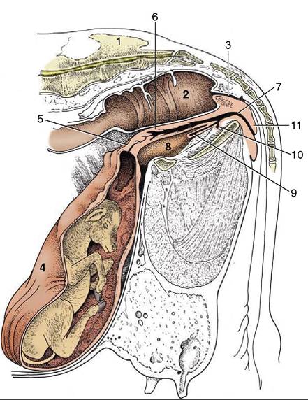
Figure 29-24 Paramedian section of the caudal abdomen and pelvis of a pregnant cow. The section is not quite vertical because it cuts through the vertebral canal and an obturator foramen. Note the large placentomes. 1, Sacrum; 2, rectum; 3, anal canal; 4, uterus; 5, cervix; 6, vagina; 7, vestibule; 8, bladder; 9, urethra; 10, suburethral diverticulum; 11, vulva.
The cervical canal is closed by a mucous plug, developing from the first month and later projecting through the external cervical ostium. The first changes in the vagina are due to traction, but the wall later becomes increasingly elastic and the lumen potentially roomier. Enlargement of the vulva is evident by the end of the first trimester in animals carrying their first calf, but in multipara, in which the vulva tends to be permanently enlarged, there may be no change obvious until shortly before birth.
Changes that signal the approach of parturition include softening of the sacrosciatic ligament, with insinking beside the tail head (Figure 29-25, A-B); a similar loosening of other pelvic ligaments allows some relaxation of the sacroiliac joints. The connective tissues of the cervix and caudal reproductive tract and vulvar and perineal skin share in these changes that, though spread over several weeks, are much intensified in the last few days. When parturition actually impends, edema of the soft parts may cause the vulva to gape.
The earlier description of the bony pelvis of cows will have suggested that it is not particularly favorable to easy parturition.
Its dimensions are relatively small, and the axis of the birth canal is broken where it passes over the pubic brim and again where the floor changes direction to rise toward the exit. Some increase in the vertical diameter is possible if the pelvis can rotate about the relaxed sacroiliac joints, but this relief is clearly denied to the standing cow. The principal soft tissue impediments to easy birth are the cervix, the caudal end of the vagina, and the vulva.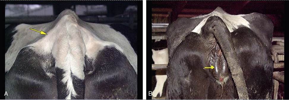
Figure 29-25 Indications of impending parturition. A, Relaxation of the sacrosciatic ligament. B, Swelling of the vulva.
Normally these parts also loosen under hormonal influence.
The umbilical cord ruptures when the cow gives birth and, being relatively short, often before delivery is complete. Its constituents part at different levels.
The fetal membranes (the “cleansings” in lay speech) normally separate from the endometrium and are expelled shortly after delivery; it is a process hastened by suckling, which stimulates the release of oxytocin. Retention with corruption in utero may require human intervention to accomplish their removal.
After parturition, the tract tends to return to its former state, but first pregnancies leave a permanent legacy in the form of thickening and loss of symmetry (Figure 29-26). The uterus contracts as soon as it empties, undergoing a very rapid atrophy in which a third of its weight is lost within a couple of days; the second third is lost before the week is out. The decline is slower thereafter, but should a cow remain “empty,” a period of superinvolution (lactation atrophy) may follow, in which the size of the uterus drops below the resting norm. Involution of the vagina, vestibule, and vulva is slower.